


As a parent or caregiver of a child diagnosed with mandibulofacial dysostosis (MFD) or Treacher Collins Syndrome (TCS), you may feel overwhelmed and anxious about your child’s future.
It’s natural to worry about the potential for bullying, discrimination, or social isolation due to their appearance. Concerns about developmental delays and the ability to lead a normal, fulfilling life can also weigh heavily on your mind.
It’s normal to feel anxious about how your loved one’s facial condition will affect their quality of life. After all, you want your child to live a happy and enriching life despite the challenges posed by MFD.
Unfortunately, navigating treatment options for MFD can be daunting. There’s just too much information to handle and too many treatment options to choose from, making it more challenging to determine how to start.
Consulting multiple healthcare providers without finding the right fit can add to the frustration and uncertainty.
You might think that these specialists don’t have the skills and resources to provide you and your child with the support you need as you go through the treatment process.
In the end, you may feel like you’re always starting over, trying to understand MFD and what treatment your child needs, no matter how much effort you’ve put in.
Dr. Youssef Tahiri, a leading global figure in craniofacial and pediatric plastic surgery, understands the unique and complex challenges you and your child experience due to MFD.
MFD manifests with noticeable features, like underdeveloped cheekbones, a small jaw, and malformed ears.
These traits don’t just affect your child’s physical appearance. They can also impact their routine bodily functions, such as breathing and swallowing.
Because MFD involves various areas of the face, treatment requires the expertise and experience of a craniofacial team and a coordinated care plan.
Our team at Tahiri Plastic Surgery is experienced in navigating the complexities of TCS and providing high-level, tailored treatments to improve your child’s well-being.
Dr. Tahiri is a world-renowned pediatric craniofacial surgeon specializing in microtia treatment. He was hand-selected by the founder of the Medpor™ technique (an innovative ear reconstruction method) to continue this practice.
Why does this fact matter to you? TCS (also known as MFD) is the most common microtia-associated syndrome.
Having an experienced and world-class microtia repair surgeon, like Dr. Tahiri, work on your child’s case is a significant advantage.
He is well-equipped to address the specific needs of MFD patients, increasing the likelihood of optimal treatment outcomes.
Are you still unsure whether to move forward with us due to the possible complexities you might encounter during the treatment process?
We understand your need to feel heard and supported, especially as you and your child navigate through the unfamiliar world of MFD.
You don’t have to go through this ordeal alone. Our professional, caring staff will guide you through every step of the journey. They will coordinate all medical appointments and aftercare, prioritizing your peace of mind.
We also offer personalized concierge services, taking care of travel arrangements, accommodations, and other logistical needs to ensure a hassle-free experience.
At Tahiri Plastic Surgery, we go the extra mile to provide cutting-edge, compassionate, customized, and comprehensive care.
We’ll manage the complexities so you can focus on what matters most —your child’s well-being.
To learn more about MFD and the treatment procedures, contact us today and schedule a consultation.
What Is Mandibulofacial Dysostosis?
If your child has mandibulofacial dysostosis, you likely see:
- Malformed or missing ears
- Underdeveloped facial bones
- Noticeably small chin and lower jaw
- Drooping eyelids
But what exactly is MFD? Simply put, MFD is a rare congenital disorder (a condition from birth) that affects the development of the face and the cranium.
As a syndrome, MFD has variable phenotypic expression, which manifests differently (meaning there is more than one identifying symptom or feature).
While children with MFD have many facial features in common, the severity of the condition varies widely.
Causes
Treacle ribosome biogenesis factor 1 (TCOF1) mutations cause MFD.
The TCOF1 gene provides the instructions for making the treacle protein, which is vital for forming bones and other tissues of the face during early embryonic development.
TCOF1’s instructions to produce treacle can be disrupted or altered due to the insertion or deletion, which are types of gene mutations, of a few DNA (deoxyribonucleic acid) building blocks.
As a result, the body makes less ribosomal RNA (rRNA), causing some cells that help develop facial bones and tissues during early development to self-destruct (also known as apoptosis).
Note: RNA (ribonucleic acid) is a molecule that helps synthesize protein.
Many researchers think that this atypical cell death might be why MFD syndrome causes developmental problems in the face.
For instance, mutations of TCOF1 in mice led to problems with the growth and movement of cranial neural crest cells into the first and second branchial arches, which are essential for head development.
Mouse models with mutated TCOF1 exhibited underdeveloped ears, facial tissues, and craniofacial bones.
That said, scientists remain unsure why the rRNA reduction usually affects the face and not other body parts.
MFD’s entire etiology (origin or cause of a disorder) is not yet fully understood. Some say MFD results from haploinsufficiency, a phenomenon in which a single normal copy of the gene is insufficient to maintain the normal cellular function of a gene.
Inheritance
While MFD is a congenital condition, it is not always inherited. In fact, about 60% of MFD cases happen randomly (de novo mutation) and have no family history.
MFD can be passed down through two inheritance patterns: autosomal dominant (the usual one) and, less often, autosomal recessive. Here’s more info about the inheritance patterns of MFD through the human chromosome:
- Autosomal dominant inheritance: If a parent has MFD, their child has a 50% chance of inheriting the TCOF1 variant (and possibly the condition).
- Autosomal recessive inheritance: Suppose the parents of a child with MFD carry one altered gene associated with MFD (POLR1C or POLR1D) but don’t show symptoms (recessive). In that case, each of the child’s siblings has:
-
- 25% chance of having MFD
- 50% chance of carrying the gene without symptoms
- 25% chance of being completely unaffected
The POLR1C (RNA polymerase I and III subunit C) and POLR1D (RNA polymerase I and III subunit D) genes are crucial to developing the structures of facial bones and tissues.
MFD symptoms vary widely within affected families. Some may have mild MFD and may go undiagnosed because their MFD-related physical traits are unnoticeable.
Meanwhile, others may have to face life-threatening risks, like airway compromise, due to severe facial problems.
If a family member has a mutated gene, affected individuals may consider prenatal (before birth) and preimplantation (the period of embryonic development between fertilization and implantation) genetic testing.
Signs And Symptoms Of Mandibulofacial Dysostosis or Treacher Collins Syndrome
MFD (mandibulofacial dysostosis) or TCS (Treacher Collins syndrome) is phenotypically heterogeneous, with different expressions or symptoms.
The major clinical features of MFD involve specific facial bones, ears, and soft tissues encircling the eyes. Here are the typical manifestations of MFD:
- Face
-
- Malar hypoplasia (underdeveloped cheekbones)
- Clefting of the zygoma, a paired, irregular bone that shapes the front and side parts of the face
- A protruding face with a receding chin and jaw, resulting in a class 2 malocclusion (overbite)
- Eyes and Eyelids
-
- Downward slanting eyes accompanied by colobomas (areas of missing tissue in the eye) of the lower eyelid
- Partial absence of eyelashes on the lower eyelids
- Hypertelorism (having widely spaced eyes)
- Vision loss associated with:
- Strabismus (crossed eyes)
- Refractive errors, such as nearsightedness or farsightedness
- Middle Ear
People with MFD might have malformed or missing ossicles—incus, malleus, and stapes—the three small bones that transmit sound waves in the middle ear.
These patients may experience conductive hearing loss, where sound waves cannot or struggle to pass through the middle ear.
This condition may require them to use a bone-conduction hearing aid for speech development.
- Inner Ear
MFD usually does not affect the inner ear.
However, the cochlea (the bony spiral organ in the inner ear) and the vestibular apparatus (the inner ear structures responsible for maintaining position and balance) of people with MFD may be malformed.
- External Ears
-
- Common occurrence of microtia, in which the external ear structures are usually small, absent, or malformed (the outer ears may be rotated or crumpled)
- Atresia (underdeveloped ear canal)
- Extra ear tags
- Stenosis (narrowing of the external ear canals)
- Small growths of skin or pits in front of the external ear (preauricular tags)
- An abnormal tube closed on one end (blind fistula) drains the ears to the nose
- Airways
People with MFD may experience potential airway restriction and respiratory issues, like obstructive sleep apnea (a condition where normal breathing and air movement are repeatedly interrupted during sleep).
Some MFD-related respiratory conditions may require interventions such as:
-
- Tracheostomy: This surgical method opens the windpipe through the neck to help air and oxygen reach the lungs.
- Mandibular distraction: This procedure lengthens a small or recessed (indented or hollow) jaw.
- Prone positioning: This technique involves placing a patient with ARDS (acute respiratory distress syndrome) face down to enhance their breathing and lung function.
Children may experience obstructive sleep apnea, characterized by repeated short interruptions of normal breathing during sleep.
- Mouth
-
- Difficulty swallowing due to craniofacial abnormalities that may require tube feeding or the insertion of a gastrostomy tube
- Possibility of cleft lip and palate
- Choanal atresia (narrowing of the back of the nasal cavity)
- Neurological
While the mental capacities of people with MFD usually remain intact, they may experience brain and behavioral issues, like microcephaly (underdeveloped head) and psychomotor delay.
According to the National Organization for Rare Disorders, as of 2023, 5% of individuals with MFD show development deficits or neurological issues.
For instance, an MFD patient may experience speech and language impediments due to cleft palate, hearing loss, or jaw and airway issues.
That said, normal language development doesn’t usually impact intelligence.
Other less common symptoms that may also be associated with MFD include:
- Congenital heart defects, such as:
- Malformed atrial and ventricular septa: This malformation may happen when there are holes between the upper and lower chambers of the patient’s heart.
- Persistent truncus arteriosus: This condition occurs when the developing truncus does not split into the aorta and pulmonary artery, leaving a single, large blood vessel exiting the heart.
- Gastrointestinal malformations, including:
- Chronic pseudo-intestinal obstruction (CIP): CIP is a rare gastrointestinal condition that affects peristalsis (the involuntary, coordinated muscular contractions of the gastrointestinal tract).
- Esophageal regurgitation: This disorder is typically associated with an abnormally relaxed LES (lower esophageal sphincter). Abnormal relaxation of the LES causes gastric content to reflux back into the esophagus.
Physical Features

Here are more detailed descriptions of the typical physical characteristics commonly associated with MFD:
- Cheeks: The cheeks of infants with MFD may appear flat or sunken due to underdeveloped or missing cheekbones.
- Jaw: MFD patients may also have micrognathia (a condition in which the mandible and the chin appear small) due to the underdeveloped structure of the lower jaw bone (mandible).
Some children may exhibit features usually associated with the Pierre Robin sequence, including severe micrognathia.
This condition happens when the tongue sits further back in the mouth than usual (glossoptosis), sometimes with incomplete closure of the roof of the mouth (cleft palate).
Also, bony structures, like coronoid and condyloid processes, anchoring the mandible to muscle are typically flat or missing.
- Teeth: Children with MFDs may also have dental problems, such as:
- Misaligned teeth (malocclusion)
- Underdeveloped (hypoplastic) teeth
- Missing teeth
- Discoloration or clouding of the teeth’s enamel
- Maxillary molars (the six molars on the upper jaw) that may erupt ectopically (which happens when the teeth don’t align according to their usual eruption pattern)
- Eyes and Eyelids: MFD may also affect the eyes’ structure and surrounding area. For example, a child with MFD may experience any or all of the following:
- Lower eyelid notch
- Narrowed tear ducts
- Crossed eyes
- Malformation of the globe of the eye due to microphthalmia (abnormally small eyes) or cleft of missing iris tissue
These abnormalities may be the reason why some MFD patients experience vision loss.
The extent of visual impairment varies depending on the severity of the ocular abnormalities and which abnormalities are present in the eye.
Other Physical Differences
Some individuals with MFD show additional (less common) physical differences, such as:
- “Beaked” nose
- Cervical spine malformation, which may result in:
- The rupture, protrusion, and bulging of the cervical disc
- The narrowing of the spinal canal around the spinal cord
- The abnormal wearing out of the cartilage and vertebrae
- Macrostomia (wide mouth)
- Unusual growth of the scalp hair down the cheeks
The symptoms and severity of MFD can vary from one affected individual to another, even among members of the same family.
Some people’s MFD symptoms may be so mild that they can go undiagnosed. Others may have noticeable abnormalities and be at risk of life-threatening respiratory complications.
Therefore, it is important to note that an MFD patient may not have all the above symptoms.
If you have a child with a noticeable congenital disorder, you may be struggling with understanding your situation.
You may have questions like, “Does my child really have mandibulofacial dysostosis?” “What does MFD look like?” and “How would the physical differences affect his life?”
While there is still more to learn about rare congenital disorders like MFD, Dr. Tahiri can help you understand and address your child’s condition.
Diagnosis
Mandibulofacial dysostosis has various symptoms similar to other craniofacial conditions.
For example, like MFD, Nager syndrome—a condition caused by the mutation of the SF3B4 (splicing factor 3B subunit 4) gene, which is responsible for producing the SAP49 protein—may feature the following characteristics:
- Malar hypoplasia
- Downward slanting of the eyes
- Microtia (ear malformation)
Spliceosome-associated protein 49 (SAP49) is part of a complex called a spliceosome, which aids in the processing of messenger ribonucleic acid (mRNA). Meanwhile, mRNA serves as the genetic blueprint for proteins.
While how your baby’s face looks at birth may cause doctors to suspect MFD as the most likely diagnosis, it is not definitive.
Your doctor may also order an X-ray imaging of your child’s facial bones and genetic tests to confirm their diagnosis.
If you have other family members with MFD, genetic testing can determine the presence of MFD mutation.
At Tahiri Plastic Surgery, we offer holistic and thorough care. We combine state-of-the-art diagnostic procedures with the highest-level care to effectively diagnose craniofacial conditions, including MFD.
We follow a meticulous approach to diagnosing MFD, including:
- Performing a thorough clinical evaluation
- Conducting an in-depth examination of your child’s medical history
- Identifying crucial physical features associated with MFD
Assessing Mandibulofacial Dysostosis with Imaging Techniques
Like in other congenital conditions, imaging can be crucial to determining an MFD diagnosis.
This assessment method lets your doctor dig deep into distinctive craniofacial features that a clinical examination might miss.
For instance, physical abnormalities, like microtia, can be a part of various syndromes, including MFD, Goldenhar syndrome (a condition affecting the development of the nose, ear, lip, and soft palate), and Nager syndrome. Here are some imaging techniques that your doctor may use to assess MFD:
- X-ray Imaging: Specialized X-ray evaluations may determine the nature and severity of MFD-associated symptoms while differentiating MFD from look-alike syndromes.
These imaging tests may show the following:
-
- Additional ear deformities that were not detected during a clinical examination
- Micrognathia (smallness of the jaw) due to the underdeveloped lower jawbone
- Presence and extent of hypoplasia in the face and skull area
X-ray imaging (coupled with a thorough clinical evaluation) can also identify the subtle manifestations of MFD in affected individuals who don’t exhibit noticeable symptoms.
Still, your doctor may recommend genetic testing to confirm the diagnosis based on the X-ray imaging.
- Fetal Ultrasonography
This imaging technique uses reflected sound waves to visualize the developing fetus and discover prenatal traits suggestive of MFD.
Suppose your child’s MFD presents a cosmetic or functional concern. In that case, it will be best to consult a certified specialist to help you assess what treatment suits your loved one’s current situation.
Tahiri Plastic Surgery has a well-rounded medical and administrative team. Our staff members support Dr. Tahiri in giving patients our best and most comprehensive care.
We are committed to providing holistic, compassionate medical care that addresses the needs of both the child and the parent.
Treatment
Like most congenital disorders, mandibulofacial dysostosis has no cure. So, MFD treatment focuses on addressing specific symptoms and needs apparent in an affected individual.
For instance, MFD care may involve early surgical interventions to resolve feeding, breathing, and structural issues from birth.
Treatment procedures for MFD vary in scope, purpose, and target areas.
While some patients may only need nonsurgical interventions, such as dental work, speech therapy, and hearing aids, others may require surgical reconstruction of craniofacial malformations.
Depending on the severity of your child’s MFD, your doctor may recommend the following treatments:
- Tracheostomy
- Glossopexy (tongue-lip adhesion)
- Mandibular distraction osteogenesis
- Gastrostomy
- Ear reconstruction
Another crucial fact about MFD is that it affects various face and skull areas. As such, an interprofessional team approach is ideal to ensure comprehensive treatment.
At Tahiri Plastic Surgery, we aim to provide patients and parents with thorough, exceptional, and holistic services that address the medical, surgical, and psychosocial aspects of MFD.
Dr. Tahiri’s personalized approach and unwavering attention to detail make us the best choice for your little one’s care.
Management Of Mandibulofacial Dysostosis
MFD is a rare, complex craniofacial disorder. It varies in extent, severity, and expression.
That’s why managing patients with MFD demands a coordinated effort from various specialists in multidisciplinary settings.
Long-term MFD management focuses on these:
- Monitoring growth and development
- Addressing new concerns
- Providing support for facing educational and social challenges
Ear Malformations and Hearing Impairment
Bilateral (affecting both sides of the face) malformations of the inner and outer ear (for example, microtia and atresia) are a typical feature of mandibulofacial dysostosis.
That’s why MFD patients may experience hearing loss of varying degrees, depending on the severity of their ear deformities.
Say your child is diagnosed with MFD and appears to experience hearing impairment. In that case, they may benefit from hearing rehabilitation, ear reconstruction, or both.
- Hearing Rehabilitation
A pediatric otolaryngologist—a doctor specializing in nose, ear, and throat treatment—may conduct formal audiological testing to gauge your child’s hearing abilities.
After the evaluation, this specialist may fit your loved one for hearing aids to help facilitate better communication.
Here are two ways specialists help MFD patients develop speech and language skills despite ear malformations:
-
- Bone conduction amplification: According to the National Institutes of Health (NIH), bone-anchored hearing aids (BAHA) stand out as superior alternatives to conventional bone-conduction hearing aids. The NIH recommends BAHA for patients above three years of age.
Meanwhile, a banded hearing aid is ideal for those younger than three. You can click https://www.ncbi.nlm.nih.gov/books/NBK562230/ to read NIH’s full content about MFD.
-
- Speech therapy: Speech pathologists may help a child with MFD improve their communication skills.
- Ear Reconstruction
People with MFD usually have microtia (outer ear malformation), specifically bilateral microtia. As such, they also often experience bilateral conductive hearing loss.
Adequate and early intervention with a banded hearing aid is the primary way to address this problem.
Now, cartilage ear reconstruction, a common external ear reconstruction procedure, is typically staged when the patient is around six to nine years of age and has adequate rib cartilage to help with the repair.
However, your child can undergo ear reconstruction as early as 3.5 years of age through the Medpor™ technique, which Dr. John F. Reinish, a world-renowned pediatric plastic surgeon, invented.
The Medpor™ ear reconstruction procedure uses a 3D (three-dimensional) porous polyethylene implant to fix ear malformations.
In 2016, Dr. Reinisch handpicked Dr. Tahiri, a highly regarded pediatric craniofacial surgeon, to join him at the forefront of microtia treatment and continue the Medpor™ practice.
As a leading surgeon in ear reconstruction, Dr. Tahiri has extensive experience in treating patients with MFD.
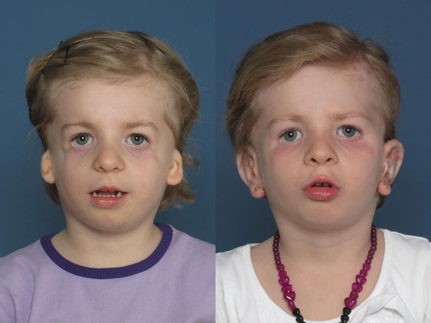
Click here to see Dr. Tahiri’s amazing work. Learn how a 4-year-old girl with MFD and bilateral microtia underwent bilateral ear reconstruction using a porous high-density polyethylene ear implant.
Obstructive Sleep Apnea
Early, coordinated care can help promptly identify obstructive sleep apnea and assess upper airway blockage levels.
Newborns may face airway-related challenges due to maxillary or mandibular hypoplasia (underdevelopment of the bones of the upper or lower jaw), potentially necessitating specialized positioning, oxygen supplementation, or tracheostomy.
While some may require prolonged tracheostomy, neonatal mandibular distraction offers an alternative.
This method involves the insertion of lengthening (distraction) devices into the jaw.
Tracheostomy is an invasive procedure that may interfere with speech and can result in scar tissue below the vocal cords and subsequent stenosis.
Conservative and surgical interventions aim to reduce its necessity. Ongoing respiratory evaluations are crucial, particularly for individuals with both MFD and cleft palate.
Feeding Difficulties
A cleft palate (an MFD symptom) may prevent babies from sucking and swallowing.
A surgeon may implant a tube into the stomach of an affected infant experiencing feeding difficulties to help them receive sufficient calories (gastrostomy).
MFD patients with micrognathia (a condition where the lower jaw is smaller than usual) or choanal atresia (wherein an excess tissue blocks the baby’s nasal airway) may also require feeding support via gastric tube.
Speech Delay or Impairment
Children with MFD may experience speech delay or impairment due to the following:
- Stenosis and subglottic scar tissues caused by tracheostomies
- Mandibular (jaw) size
- Occlusal discrepancies (poor alignment of teeth)
- Hypernasality (too much sound vibrates in the nasal cavity during speech) and hyponasality (not enough sound resonating in the nose during speech) caused by choanal atresia
At the outset, focusing on restoring hearing and speech promptly and facilitating the timely initiation of speech therapy can help address speech-related problems.
Eye Anomalies and Visual Impairment
Fixing uneven eye position with surgical procedures is rarely necessary and doesn’t always result in a satisfying look.
Typically, the position of the MFD patient’s eyes is due to their skull and face shape and doesn’t usually cause any functional issues. So, even if their eyes aren’t quite in the right place, their vision may still be fine.
However, if your child has colobomas (holes in their eyes) or their eyelids don’t adequately cover their eyes, it’s important to get help early on to protect their vision.
In really severe cases, surgery might be needed to lengthen your eyelids. For instance, a detailed reconstruction approach may fix lower eyelid colobomas.
The initial corrective surgery usually happens around age six or seven. This procedure aims to fix the antimongoloid slant of the palpebral fissures (the area between the open eyelids), a common feature of MFD.
It involves correcting both the bone structure and the underdeveloped lower eyelid (hypoplasia). Surgeons use a specific approach that involves making a small incision under the malformed eyelid to ensure thorough repair of the zygomatic-malar (cheekbone-related) defect.
Malar and Mandibular Hypoplasia
Some MFD patients may require craniofacial reconstruction. The timing of the procedure usually depends on the individual’s needs, preferences, and severity of symptoms.
That said, the best aesthetic results usually come when these surgeries are performed near the time of early skeletal maturity (17 to 21 years old in males and 15 to 17 years old in females).
Jaw correction may be considered from ages 13 to 16, depending on the condition and development of the temporomandibular joint (the joint that connects the skull and the lower jaw).
As patients approach skeletal maturity (around age 16), they can have their facial soft tissue refined using additional fat grafting.
These cosmetic and functional enhancements from craniofacial reconstruction can significantly affect patients’ feelings about themselves and their interactions with others.
A study involving 20 MFD patients reported that those who underwent surgery had a noticeable improvement in their self-esteem, appearance, and functional ability, especially a year after the operation.
Still, there are no set treatment plans for patients with MFD, so determining when to undergo craniofacial procedures can be challenging. For example:
- Reconstructive surgeries on the eye sockets (orbitals) and cheekbones (zygoma) may be completed around five to seven years old.
- Orthognathic (related to the jaw and the associated malocclusion or misalignment of teeth) therapies are typically completed before 16 years of age.
- Nasal reconstruction (septorhinoplasty), if deemed necessary, is usually postponed until after the orthognathic procedures, usually after the age of 17.
Cleft Palate
Cleft lip and palate are usually surgically corrected at 3 and 10 months of age, respectively.
Before performing the surgery to close the cleft palate, doctors may conduct a sleep study to simulate the effects of the closure. This method helps them prevent any potential breathing difficulties that could happen post-surgery.
Psychological Concerns
Aside from the physical and medical challenges, mandibulofacial dysostosis can have profound psychosocial implications.
MFD patients may experience psychological distress and face social challenges, like bullying, stigma, and prejudice because they look and act differently.
Even you as a parent may also bear the psychological burden due to MFD. You may have had to endure judgmental stares, unsolicited advice, and derisive comments due to your child’s condition.
Dr. Youssef Tahiri finds it very hard to see families and children who can’t get care.
He founded Tahiri Plastic Surgery to ensure parents and children with MFD can get the compassionate and comprehensive treatment they need and deserve.
As such, our practice is committed to building a supportive community comprising family, educators, healthcare providers, and peer support groups to nurture self-esteem, independence, and resilience.
We aim to help affected individuals and families build confidence and lead normal lives.
How Common Is Mandibulofacial Dysostosis?
The occurrence of mandibulofacial dysostosis may range from 1 in 25,000 to 1 in 70,000 live births, with the most commonly reported figure being 1 in 50,000.
There is no racial or gender preference when it comes to MFD. Also, the severity of the malformation the patient is born with tends to stay the same and does not worsen over time.
Other Names For The MFD Condition
MFD is entry #154500 in the OMIM (Online Mendelian Inheritance in Man), a well-known catalog of human genes and genetic disorders.
You may also know MFD as:
- Berry’s syndrome: MFD was named this due to its early recognition by the ophthalmologist Dr. George Andreas Berry in 1889.
- Treacher Collins syndrome: Edward Treacher Collins may have been the first person to identify the chief feature of MFD, namely, malar bones’ marked hypoplasia (underdevelopment).
This underdevelopment usually gives the patient’s cheeks a flattened appearance.
- Franceschetti–Klein syndrome: Adolphe Franceschetti and David Klein further described the facial features of the condition and coined the term mandibulofacial dysostosis.
Some use MFD and MFDM (mandibulofacial dysostosis with microcephaly) interchangeably. However, these conditions arise from different mutations.
MFD may be caused by mutations of the TCOF1 (treacle ribosome biogenesis factor 1), POLR1D (RNA polymerase I and III subunit D), and POLR1C (RNA polymerase I and III subunit C) genes. In contrast, MFDM occurs due to EFTUD2 (elongation factor Tu GTP binding domain containing 2) gene mutations.
EFTUD2 is a gene that contains the instructions for making one part of two complexes known as the major and minor spliceosomes.
Here are other notable figures who contributed to the literature about MFD:
- Michael Dixon, PhD, University of Manchester, United Kingdom
- Paul Trainor, PhD, Stowers Institute for Medical Research
- Ethylin Wang Jabs, MD, Department of Genetics and Genomic Sciences, Mount Sinai School of Medicine
How Long Do Kids With Mandibulofacial Dysostosis Live? What Is The Life Expectancy Of Someone With MFDM Syndrome?
The prognosis for individuals with mandibulofacial dysostosis varies depending on the extent of the abnormalities and the success of the interventions.
Many people with MFD lead full, active lives, usually with the help of ongoing medical care.
Generally, MFD patients with access to proper management have a normal life expectancy.