Medically reviewed by Youssef Tahiri, M.D.
As a parent, you want to give your child the best chance at a happy, fulfilling life. You hope they’ll enjoy their childhood and, as they grow, discover new joys through hobbies and lasting friendships.
Hearing that your child has a congenital condition like microtia and atresia can be overwhelming. Questions might flood your mind, such as:
The good news is that hope is within reach.
At The Los Angeles Ear and Craniofacial Center, led by Dr. Youssef Tahiri, you’ll find a trusted partner to help you navigate these challenges. Here, you can ask tough questions and receive clear, honest answers—paired with solutions tailored to your child’s needs.
Dr. Youssef Tahiri deeply understands what you’re going through. He has helped many families through the complexities of microtia and atresia with expertise and compassion.
As a leading pediatric and adult plastic and craniofacial surgeon, Dr. Tahiri creates personalized treatment plans to fit each patient’s unique needs and situation. His approach prioritizes minimizing pain, reducing scarring, and, in some cases, offering outpatient procedures.
Whether through reconstructive surgery, hearing restoration, or advanced 3D ear reconstruction techniques, we’re dedicated to helping your child thrive. Our goal is to improve both their appearance and hearing, giving them the confidence to embrace life fully.
Innovative options like the PPE (porous polyethylene) ear reconstruction technique and aural atresia repair provide real hope for a brighter future—free from constant worry.
Learn more about how we can help you and your child overcome the challenges of microtia and atresia.
Let’s start with the basics: Microtia is a rare congenital condition affecting the outer ear (pinna).
If your child was born with a small, underdeveloped, or missing ear, they may have microtia.
This condition is often linked with aural atresia (the absence of an external auditory canal) or canal stenosis (extremely narrow ear canal). As a result, children with microtia typically experience hearing challenges.
Although microtia is uncommon, it still impacts many lives. Approximately 1 to 5 out of every 10,000 births involve this condition.
If you’re learning about microtia for the first time, it’s natural to feel overwhelmed. But take heart—there’s good news. You and your child don’t have to face this journey alone. With the right care and support, your child can live a full, happy, and confident life.
At The Los Angeles Ear and Craniofacial Center, we specialize in providing expert, personalized, and compassionate patient care for children and adults with microtia.
Dr. Youssef Tahiri and his dedicated team work to make the treatment process as smooth and stress-free as possible for both you and your child.
“Dr. Tahiri is an amazing surgeon. We have known him for over five years, and he has been supportive over the years. He came to numerous microtia and atresia events that our family helped host. He patiently walked us through the entire surgical process and always answered our questions. My son had ear reconstruction surgery two months ago and also had the Oticon Ponto abutment procedure. We are beyond thankful for Dr. Tahiri, [and] his team, especially Paola and the K & B Surgery Center. They all together took care of our son like their own. We would recommend Dr. Tahiri again and again to all prospective patients. We have met multiple surgeons, and we knew he was the one the moment we met all those years ago.”
Christine Bayan
“My son had a [porous polyethylene] ear by Dr. Tahiri, and he is an amazing doctor [who] took the best care of my son! His staff is great to work with as well.”
Veronica S.
Microtia manifests in different ways, ranging from mild anatomical issues to the total absence of the external ear. For this reason, the medical community categorizes microtia into four types, each marked by distinctive physical differences:

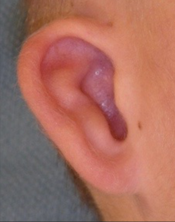
All parts of the outer ear are present, but the external ear’s size appears smaller than usual. Individuals with type 1 microtia may have a missing or narrowed ear canal.

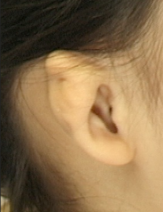
The external ear is small and underdeveloped (especially the top two-thirds of the outer ear). Patients with type 2 microtia often have narrowed or missing ear canal.

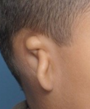
The ear’s cartilaginous framework, including helix, antihelix, and concha, is underdeveloped. The only part formed is a lobule, a small piece of the ear lobe (the soft, fleshy part at the bottom of the outer ear).
Type 3 microtia is the most common grade of microtia.

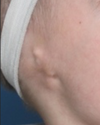
External ear structures are completely absent (anotia). Patients with type 4 microtia have a low-set hairline and a missing ear canal.
In addition to these four categories, microtia ears may be classified into lobular type (those with a prominent lobule remnant) and conchal type (often associated with a narrowed canal).
Each microtia category presents unique challenges that only experienced ear reconstruction specialists like Dr. Tahiri can accurately identify and effectively address.
The CDC (Centers for Disease Control and Prevention) defines disability as any condition that limits a person’s activity and capacity to interact with the world.
For instance, a disability may make it difficult for someone to hear, see, think, or move.
Based on this definition, microtia itself isn’t a functional disability. That said, hearing loss, which is often associated with microtia, can limit the affected individual’s activity and social participation.
As such, hearing loss may qualify a microtia patient for disability benefits, depending on the condition’s impact on their life.
Does your child struggle with life-impacting symptoms due to microtia? If so, getting comprehensive care is vital to enhance their quality of life:
Ideally, bone conduction hearing aids should be fitted before your child reaches six months of age so they can catch the sounds they need to develop their speech and language skills.
At The Los Angeles Ear and Craniofacial Center, we partner with you to create a treatment plan tailored to your needs. Dr. Tahiri is deeply committed to helping families manage microtia and its related conductive hearing loss. This means sounds are blocked before they reach the inner ear.
If you or your child have microtia and want to understand your options, Dr. Tahiri encourages you to schedule a video consultation.
For those in Los Angeles, in-person consultations are available to provide more personalized care and guidance.
To ensure a comprehensive clinical evaluation, pediatric doctors and otolaryngology specialists must address audiological and aesthetic concerns and other microtia-associated issues.
People with microtia might also have to face these malformations:
Also known as Goldenhar syndrome (GS), HFM is characterized by three unique features:
For the ears, patients may experience:
Do you worry these deformities will prevent your child from leading a regular life? Don’t be—you’re not alone.
Dr. Tahiri specializes in various surgeries, from microtia repair to skull reshaping and jaw realignment.
He can restore balance and harmony to your child’s face with care and precision.
VPI occurs when the muscle separating your mouth and nose (velopharyngeal sphincter) doesn’t close completely.
This condition can cause speech to sound nasal and, in severe cases, food or drinks to pass through the nose.
Dr. Tahiri diagnoses VPI by conducting a clinical test, speech evaluation, and nasopharyngoscopy (a procedure that uses a small camera to view the throat and nasal cavity).
VPI treatment typically involves surgery to correct air leakage and hypernasal speech. That said, not all patients have to undergo surgical correction.
As an experienced facial plastic and reconstructive surgeon, Dr. Tahiri can determine the optimal approach to each VPI case.
Untreated microtia can negatively impact an individual’s development, especially if the condition causes the person to experience hearing loss.
For example, hearing issues can lead to speech delays, affecting school performance and development.
These challenges don’t have to define your child’s future. With early diagnosis and expert care, your little one can still live a fulfilling life.
That’s where Dr. Tahiri steps in. He offers a comprehensive approach, coordinating with audiologists, otologists, and other specialists to ensure every aspect of your child’s health is addressed.
Dr. Tahiri aims to address all microtia-related concerns—physical appearance, hearing, and psychosocial health. From the time you contact us, we’ll guide you and your family, beginning with the reassurance that you’re not at fault for your child’s condition.
Once you’ve clearly understood the condition and your treatment options, we’ll partner with you to determine the proper care for your child.
People with microtia (malformed outer ear) often also have aural atresia (underdevelopment or absence of the ear canal).
Like microtia, atresia can be categorized according to its unique clinical presentations:
These physical abnormalities may affect only one ear (unilateral atresia) or both ears (bilateral atresia).
Atresia can also be acquired and congenital (something you’re born with). Acquired atresia may result from infections, inflammation, accidental trauma, radiation therapy, and benign or malignant tumors.
Meanwhile, the cause of congenital aural atresia has yet to be fully understood, but it seems to occur when branchial arches don’t develop properly during fetal growth.
This disruption in the development causes an otherwise healthy fetus’ lower ears, face, and neck to be malformed or underdeveloped.
Whether congenital or acquired, atresia can result in profound conductive hearing loss. If left untreated, these hearing difficulties can persist throughout your child’s life and, in some cases, may delay language development and cause learning disabilities.
That’s why early intervention—whether medical or surgical—matters. It gives your child the best shot at clear hearing and a healthy, thriving life.
If you’ve just learned that your child has atresia, you may feel overwhelmed, confused, and worried about what comes next.
Will your child completely lose their ability to hear? If so, how will they communicate and develop normally? Can aural atresia be treated or managed effectively?
Dr. Tahiri specializes in helping children and families navigate these concerns. He offers a solution that lets your child grow with confidence.
Dr. Tahiri can conduct surgery on patients as young as three years of age, allowing for healthy development and preventing future bullying.
While this case rarely occurs, microtia without atresia is possible. The only known condition in which this happens is chromosome 18q syndrome.
This genetic disorder results from a missing piece of the longest arm (called the “q” arm) of chromosome 18.
People with chromosome 18q syndrome may have narrow ear canals (stenosis) or no ear canal at all (atresia). Still, they won’t have the usual underdevelopment of the ear seen in microtia.
In some cases, their outer ears may look flat or unusually prominent.
If you’re concerned that your child may have chromosome 18q syndrome, it can be confirmed with a simple blood test.
Aural atresia and ear canal stenosis prevent sound from reaching the inner ear properly, making it difficult for people to hear.
Imagine trying to hear a conversation while wearing earplugs. You can catch bits and pieces, but the words don’t come through clearly. That’s likely how people with aural atresia hear things.
As such, children with aural atresia and stenosis struggle to understand speech, especially in noisy places.
They may also struggle to determine where sounds are coming from. For instance, they could have trouble telling if someone is speaking from behind or the side. This difficulty grows even more if both ears are affected.
Managing aural atresia and the accompanying hearing issues can be complex. It may require a team of specialists to address these conditions, including:
Dr. Tahiri understands the value of a multi-disciplinary approach when treating atresia.
That’s why he partners closely with trusted experts like Dr. Roberson, a skilled otologist who performs ear canal reconstruction (canaloplasty) to help restore hearing. They collaborate to enhance hearing and, when possible, create a new ear canal to restore the ear’s function.
Dr. Roberson reviews the actual CT scan (computed tomography scan) and uses the Jahrsdoerfer 10-point grading scale to evaluate surgical candidacy.
Treating hearing loss—whether with hearing aids, implants, or canal reconstruction—requires coordination between the ear specialist and the surgeon working on the outer ear.
Atresia repair (fixing the ear canal) usually happens after costal cartilage ear reconstruction (using rib cartilage to build the ear).
Most otologists access the ear canal through a small cut behind the ear to reach the temporal bone (the part of the skull that holds the ear).
If surgery isn’t ideal for your child, don’t worry—there are many other options to improve their hearing.
For instance, bone-anchored hearing aids (BAHA) are a popular choice, and there are versions for all ages, including a soft-band BAHA that can be worn right from infancy. Another option is the Vibrant Soundbridge, an implantable hearing device that helps boost sound directly.
Some say microtia and atresia can be linked to genetic and environmental factors.
In some cases, microtia runs in the family and is passed down through Mendelian inheritance (meaning it’s linked to genes and can follow a dominant or recessive pattern).
Other times, chromosomal abnormalities (changes in the structure of chromosomes) can cause these conditions. While researchers have identified several genes responsible for the condition, most are homeobox genes (genes that help control the body’s basic development).
That said, the exact cause of microtia and atresia is still unknown. Still, microtia and atresia can occur alone or with other syndromes that lead to malformations of the head and neck, such as Goldenhar syndrome and Treacher-Collins syndrome.
The bottom line is, as a parent, you need to know there’s nothing you could have done differently during your pregnancy that would’ve prevented your child’s condition. It’s simply not something within your control.
Dr. Tahiri offers some of the most advanced and effective solutions for managing microtia and atresia. His approach combines state-of-the-art technology with a deep understanding of cosmetic restoration and improving hearing.
For microtia, one of the standout options is the use of porous polyethylene ear implants—including MEDPOR™, OMNIPORE™, and the latest SUPOR™. These implants are made from a high-density, porous biomaterial that mimics the shape and projection of a normal ear.
As such, they allow for reconstruction using your child’s own tissue, creating a more natural, personalized look.
For atresia, Dr. Tahiri works with Dr. Roberson, a highly regarded otologist (Dr. Roberson) who will perform ear canal reconstruction (canalplasty).
With these combined techniques, Dr. Tahiri is transforming the treatment of microtia and atresia, restoring not just the ear but also the confidence and quality of life for children and their families.
Hearing loss is a common concern among children with microtia and aural atresia. The good news is that early diagnosis and treatment can make a significant difference.
Typically, hearing loss in these children is conductive due to issues with the outer ear or the ear canal. This unfortunate situation can be caused by aural atresia and Eustachian tube dysfunction (issues with the tube that helps equalize pressure in the ear).
While sensorineural hearing loss (which comes from issues in the inner ear) is less common, it can occur in some cases due to abnormalities in the inner ear, especially with conditions like craniofacial microsomia (a genetic disorder that can affect ear development).
At The Los Angeles Ear and Craniofacial Center, hearing loss treatment and ear reconstruction are always a team effort. An otologist will work with the surgeon handling the ear reconstruction to ensure both issues are addressed together.
In the past, atresia repair (fixing the ear canal) was done after ear reconstruction since most surgeons access the ear canal through a cut behind the ear (called a posterior mastoid incision). The downside of this approach is that it can interfere with the blood flow to the tissue used to cover the ear.
However, Dr. Tahiri’s approach uses an alloplastic ear implant (a synthetic ear structure) to reconstruct the ear canal before or during the ear reconstruction.
This technique is a game-changer because the canal helps position the reconstructed ear properly. Early atresia repair has the added benefit of potentially improving hearing at a critical stage in brain development.
For children who are not candidates for a functional ear canal, Dr. Tahiri can create a “faux-canal” (a fake ear canal) during the microtia reconstruction.
This involves carefully reshaping the ear’s structure, removing any leftover cartilage, and adding extra tissue to deepen the ear’s shape.
The tragus (the small bump outside the ear canal) is then crafted using tissue from the mastoid (the area behind the ear), ensuring a natural look and feel.
Dr. Tahiri is at the forefront of these advanced treatment options, always focused on what’s best for your child.
If you have questions or want to explore the best solution for your child, don’t hesitate to reach out. Your child’s well-being and confidence are his top priorities.
People with microtia typically have normal cochlear function, meaning they’re not deaf—they can hear through bone conduction (sound transmission through the skull). Normal air conduction allows hearing at 15 to 20 decibels (dB, a unit to measure sound level).
Unfortunately, for many individuals with microtia and aural atresia, their hearing loss falls in the 60dB to 70dB range, which is substantial. To put it in perspective, blocking a healthy ear canal with your finger typically reduces hearing by about 30dB.
Since normal speech is around 40 to 45dB, these children struggle to hear typical conversations.
For kids with unilateral microtia (one affected ear), the hearing loss on that side is significant. Still, they can usually hear just fine out of the unaffected ear.
As a result, speech development tends to be normal. However, things get trickier for kids with bilateral microtia (both ears affected) or if the unaffected ear also has hearing problems that go unnoticed.
The challenge with unilateral hearing loss is that it often flies under the radar. Parents of children with one microtic ear usually don’t notice any issues in their child’s early years.
Kids with unilateral microtia often develop normally, and hearing seems fine because the “good” ear compensates.
Unfortunately, as people age and start engaging in more complex conversations and environments, the gaps in their hearing become more apparent.
It’s not uncommon for children with microtia to turn their good ears toward a sound, trying to catch it better. They do so because the head itself creates a sound shadow that makes it more challenging for them to understand speech from the other side.
That’s why hearing tests, early diagnosis, and treatment are vital.
Dr. Tahiri works closely with skilled otologists to ensure your child’s hearing is addressed alongside ear reconstruction. This coordinated approach ensures that the cosmetic and functional aspects of the ear are handled with care.
Hearing devices are essential for boosting sound and improving the quality of life for those with hearing loss. From traditional hearing aids to more advanced options, they help ensure no moment goes unheard—from talking with family to enjoying everyday sounds.
At The Los Angeles Ear and Craniofacial Center, we offer Cochlear™ Osia® (Osia) placement. The Osia is a bone-conduction hearing system created by Cochlear Corporation.
Unlike a traditional hearing aid, the Osia implant has two parts: a sound processor worn behind the ear and a surgically implanted component placed under the skin behind the ear.
This cochlear implant bypasses the outer and middle ear, directly transmitting sound to the inner ear via bone conduction.
Dr. Tahiri offers Osia because it sets the bar for sound quality in conductive hearing devices. It’s built to eliminate feedback when touched, connects effortlessly via a magnet, and—here’s the big one—it’s waterproof.
That’s a huge upgrade for kids with bilateral hearing loss, who can be virtually deaf without proper hearing aids. With the Osia, they don’t just regain normal hearing—they get a chance to live better, playing and swimming without worrying about their device.
Every child’s needs regarding hearing devices are unique. Still, with the right support, they can thrive and overcome the challenges of having a small ear or no ear at all.


After several years of experience with canal surgery first followed by MEDPOR™ as a second surgery, Dr. Roberson, in cooperation with Dr. Youssef Tahiri became the only surgeon in the world performing Combined Atresia Microtia Repair (CAM) surgery. The three surgeons combined two previously separate fields of surgical practice into one unified surgery. This eight hour, outpatient procedure combining both the atresia repair canalplasty and the MEDPOR™ outer ear reconstruction. At this time, they remain the only ones performing this surgery in the world.
Having completed over hundreds CAM surgeries with Dr. Tahiri, it is commonplace for patients to select this option for Atresia and Microtia repair. Surgeries are performed in Palo Alto. The recovery time required in California following the procedure is four weeks, with post op appointments split between Los Angeles and Palo Alto.
The CAM procedure offers several advantages over traditional techniques and comes with drawbacks as well, but it is important to know that we have found no difference in hearing results in patients undergoing separate vs. CAM procedures.
The advantages of a Combined Atresia Microtia Reconstruction include:
The disadvantages of a Combined Atresia Microtia Reconstruction include:
In the evaluation process, several factors are taken into consideration when Dr. Tahiri is recommending the method of surgery that would be advised for each child. Specific case-by-case evaluation is important, as there are times where separate procedures may be recommended over the combined approach and vice versa.
CONTACT TAHIRI PLASTIC SURGERY TODAY
To find out if your child qualifies for surgery, contact our office online today. You can also call (424) 415-8881 to speak to a member of our multilingual team.
Leaving microtia untreated isn’t just about letting a visible difference persist—it also affects a child’s development and self-esteem as they grow.
The earlier you address microtia, the less it impacts your child’s development. Your child can enjoy a happy childhood, free from the social and emotional burden of having a visible ear difference.
One of the most effective treatments for microtia is biocompatible PPE (porous polyethylene) ear reconstruction. This method has been used since 1991 and has advanced dramatically since then.
Over the years, the surgery has been refined to offer better results and a smoother overall experience for the child and their family.
Biocompatible Polyethylene Implant Ear Reconstruction
3D polyethylene implants offer several significant advantages. First, the entire procedure is completed in one stage, meaning multiple surgeries are unnecessary. Your child can see the final results after just one surgery, unlike other methods that require two separate procedures.
Another major benefit is that this is an outpatient procedure (meaning your child doesn’t need to stay overnight at the hospital), and they can go home the same day.
The surgery is minimally painful, and most kids experience little to no discomfort afterward. It also leaves minimal scarring, which is a plus for cosmetic results.
The surgery can be done as early as 3.5 years of age, allowing children to develop well and avoid bullying at school or other psychosocial issues.
3D Polyethylene Ear Reconstruction
In recent years, 3D templates (three-dimensional models) have replaced the older 2D templates (two-dimensional models) as the go-to guides for microtia ear reconstruction.
One primary advantage of using 3D polyethylene ear implants is that they eliminate the risk of implant fractures, which could happen with older, less durable materials.
These 3D implants are also biocompatible, meaning they’re made from a safe material for the body. The implant has a porous structure (tiny holes), which allows the body’s cells and blood vessels to grow into the material, essentially becoming a part of the ear.
For many years, the rib cartilage method was the go-to approach for ear reconstruction in microtia cases. Unfortunately, this technique often left much to be desired regarding the final result.
Plus, children had to endure long surgeries, multiple stages of treatment, significant physical pain, and psychological stress.
These procedures were usually delayed until the child was 10 or older, requiring hospital stays and sometimes leaving them vulnerable to bullying.
That’s when a game-changing technique was introduced: PPE ear reconstruction. This method is a single-stage procedure—meaning the entire ear reconstruction happens in just one surgery.
It can also be done as early as 3.5 years old, so there’s no waiting. This method is quicker, less painful, and done on an outpatient basis—your child goes home the same day with less stress and a faster recovery.
The rib cartilage reconstruction procedure involves multiple surgeries—usually two to five—and can take years to complete.
The process can begin when a child is between 6 and 10 years old, as they’ve grown enough to provide rib cartilage for shaping an adult-sized ear. The cartilage is removed through a chest incision, sculpted into an ear framework, and placed under the scalp to heal.
The traditional method uses rib cartilage to build a new ear. While this approach has improved over the years, it still requires multiple surgeries and a significant amount of harvested cartilage, which often means waiting until the child is 10 or older.
That makes the process more challenging—not just physically, but emotionally—for both kids and parents. And if the final result doesn’t meet expectations, the whole experience can feel discouraging.
Fortunately, Dr. Tahiri offers a better alternative: using an alloplastic framework (a synthetic ear structure) covered by a thin layer of temporoparietal fascia (a type of connective tissue).
This method has several advantages. For one, ear growth slows down after about 3.5 years, so we can safely perform the reconstruction at a younger age without costal cartilage.
This scenario means the procedure can be done sooner, with fewer stages and less discomfort.
Instead of multiple surgeries, Dr. Tahiri can perform the entire reconstruction in a single outpatient procedure—often before your child starts school. The result? Less stress for the whole family, minimal downtime, and better ear definition and projection.
Another big benefit is that soft tissue coverage is vital to a successful outcome. Dr. Tahiri removes the superficial temporal fascia from under the scalp with no additional incisions.
This technique avoids the risk of scalp alopecia (hair loss) while still giving the implant a strong, healthy layer of tissue. Dr. Tahiri is highly skilled in this procedure, using advanced tools like high-power magnification and specialized retractors to ensure precision.
The most challenging aspect of alloplastic reconstruction is ensuring proper soft tissue coverage.
However, as long as a healthy fascia flap covers the implant without tension, an alloplastic reconstruction can be performed with minimal complications.
The alloplastic framework is a far cry from the old, more invasive methods. With Dr. Tahiri’s expertise, your child can have a functional, natural-looking ear with less hassle, fewer stages, and a quicker recovery.
Many scientists believe that genetics influences microtia and aural atresia. This theory is based on studies on people and research on animals with specific genetic mutations associated with the condition.
In human cases, the chances of microtia running in families vary widely, with studies showing anywhere from 3% to 34% of cases having a family connection.
Certain chromosomal regions have been directly linked to microtia and atresia:
Microtia (underdeveloped outer ear) likely stems from a problem in the neural crest cells (NCC). These cells help form the ear structures and face during development.
While the exact cause is still unclear, disruptions in how these cells behave are thought to lead to different types or grades of microtia.
They identified a small missing segment of DNA—called a microdeletion—in a region of chromosome 18 (18q22.3-q23) containing a gene known as TSHZ1.
However, the genetic picture isn’t straightforward. In a larger group of patients with other forms of CAA (congenital aural atresia)—some also had microtia (underdeveloped outer ears) or anotia (missing outer ears)—no TSHZ1 mutations were found.
This suggests that atresia can result from different genetic causes, known as genetic heterogeneity.
In short, while TSHZ1 mutations are linked to certain types of atresia, other genes are likely responsible for this condition.
Suppose your child has been diagnosed with microtia/atresia. In that case, it’s essential to understand the full scope of their condition and the treatment options available.
Schedule a consultation with Dr. Tahiri today to learn more about microtia and atresia treatment and how it could benefit you or your child.
You can call us at (310) 255-4476 or visit us at 9033 Wilshire Blvd, Suite 200, Beverly Hills, California, USA 90211.