Marginal Mandibular Nerve Palsy
If you or someone you know is dealing with marginal mandibular nerve palsy (MMNP), you already understand how challenging it can be.
The noticeable facial asymmetry, difficulty showing expressions, speech impairment, dental issues, and trouble eating and drinking can all take a toll on daily life.
And when the cause or severity isn’t clear, it’s completely natural to start searching for answers and trying to make sense of what’s happening.
You deserve clarity, an accurate diagnosis, and a surgeon who understands both the anatomy and the emotional weight that comes with facial imbalance.
That’s where Dr. Youssef Tahiri’s expertise becomes invaluable.
Recognized & Trusted By





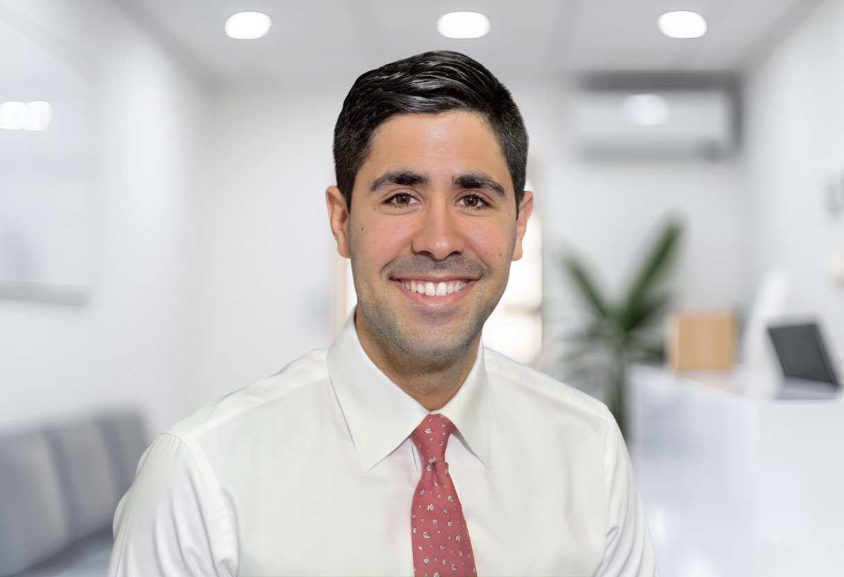
Meet Dr. Tahiri
His background includes:
- Board certification in plastic and reconstructive surgery with a subspecialty in craniofacial surgery
- Plastic and Reconstructive Surgery residency and comprehensive plastic surgery training at McGill University
- A prestigious craniofacial fellowship and Craniofacial Surgery training at the Children’s Hospital of Philadelphia
- Experience treating more than 25,000 patients from over 65 countries
- A reputation for honest communication, compassion, and caring for patients like family
If your symptoms began after surgery, trauma, or a medical procedure, you may be wondering whether the nerve is simply bruised or more seriously injured.
It’s also common to feel overwhelmed by conflicting information online or unsure about what steps to take next.
At International Microtia Institute, you’ll receive a thoughtful and detailed evaluation that identifies the source of the nerve injury and explains what’s happening in clear, understandable language.
From there, Dr. Tahiri walks you through tailored treatment options— ranging from nonsurgical balancing procedures to advanced reconstructive techniques designed to restore natural, symmetrical movement.
With experienced guidance and a clear path forward, you can approach your recovery with confidence and a deeper understanding of what’s possible.
At A Glance
Key Takeaways
A clinical summary of marginal mandibular nerve palsy (MMNP) — what it is, why it happens, and how Dr. Tahiri treats it.
-
01 Definition
Marginal mandibular nerve palsy (MMNP) is a condition affecting the ramus (branch) of the facial nerve that controls your lower lip. The condition can occur on one side of the face (unilateral MMNP) or on both sides (bilateral MMNP).
-
02 Cause
MMNP often results from surgery, trauma, or iatrogenic injury during operations in the mandibular and parotid regions.
-
03 Presentation
When MMNP is unilateral, it can cause noticeable asymmetry or weakness around the lips and mouth. Common signs include drooping of the lower lip, a crooked smile, difficulty showing expressions, or mild challenges with speaking, eating, or drinking.
-
04 Risk Factor
Conditions like microtia and hemifacial microsomia can lead to unilateral MMNP, restricting movement of the lower lip on one side while the rest of the face looks balanced.
-
05 Treatment
Dr. Youssef Tahiri offers nonsurgical and surgical treatment options for MMNP, including BOTOX®, selective myomectomy, and selective neurotomy.
-
06 Expertise
Dr. Youssef Tahiri has treated over 25,000 patients from more than 65 countries, providing expert diagnosis, personalized treatment plans, and compassionate guidance throughout recovery.
Finding Hope
What Is Marginal Mandibular Nerve Palsy?
Marginal mandibular nerve palsy (MMNP) occurs when the marginal mandibular branch of the facial nerve becomes damaged or doesn’t function properly.
This small but essential nerve runs along the lower border of your mandible. It innervates (powers) the muscles that control your lower lip— primarily the depressor anguli oris (DAO) and depressor labii inferioris (DLI).
These two muscles help pull the lower face downward during expressions, such as sadness or frowning: the DAO draws the corner of the mouth, while the DLI pulls the lower lip down.
For this reason, people with MMNP may notice lower lip weakness, asymmetry, or difficulty creating a full, balanced smile.
When MMNP affects only one side, it’s known as unilateral MMNP. During smiling or speech, the unaffected corner of the mouth drops, while the paralyzed side stays elevated, resulting in a crooked or uneven smile.
Unilateral MMNP is particularly common in patients with microtia and hemifacial microsomia, where developmental asymmetry often includes an incomplete or absent marginal mandibular nerve.
Dr. Youssef Tahiri is internationally recognized for his work in microtia, hemifacial microsomia, and craniofacial reconstruction.
With patients from more than 65 countries, Dr. Tahiri is a leading authority in restoring natural lower lip movement and achieving a more symmetric, confident smile.
Expert Insights
Understanding Marginal Mandibular Nerve Palsy
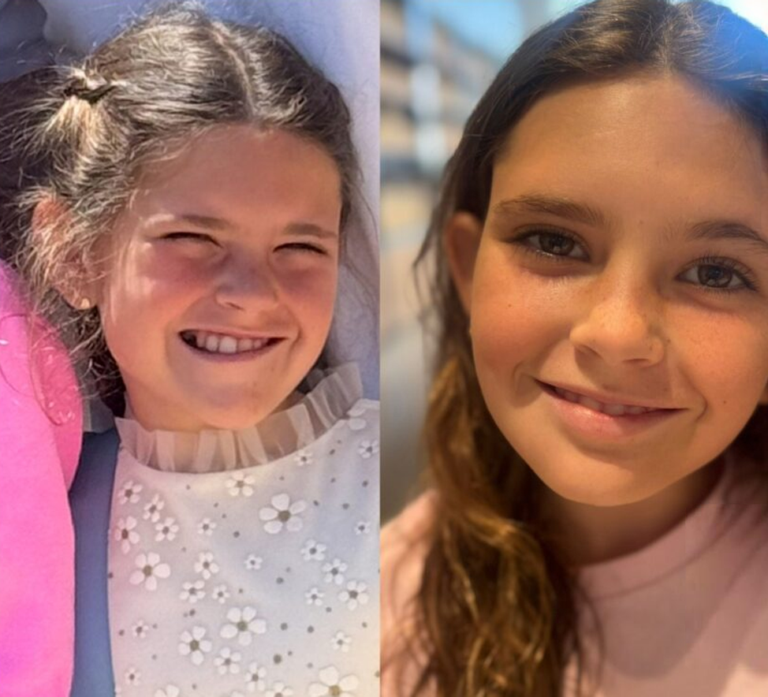
One of our happy patients before/after
A note from Dr. Tahiri & the care team
A lower lip that won’t quite move the way it used to, or a smile that suddenly looks uneven, brings up a lot of questions — and often some worry that it’s Bell’s palsy. Below is what we walk patients through in that first conversation: what this nerve actually controls, and why marginal mandibular nerve palsy is a different, more localized issue. We’ve put it here in the same words, so you have it before your visit.
What Does the Marginal Mandibular Nerve Do?
The marginal mandibular nerve controls the muscles responsible for pulling down your lower lip—especially the depressor anguli oris and depressor labii inferioris.
These muscles are essential for facial expressions like frowning, speaking clearly, and keeping your lips sealed while eating.
Normally, both corners of your lower lip move symmetrically when you smile, speak, or express emotion.
If the nerve is injured, one side of your lower lip may droop, your smile may appear crooked, and your ability to articulate words or close your mouth fully may be affected.
Understanding how this nerve functions helps you and your surgeon better identify the severity of the facial nerve injury and plan the most effective treatment path.
How Is This Different from Bell’s Palsy and Other Facial Nerve Problems?
Bell’s palsy involves sudden paralysis of the facial muscles, typically affecting one side of the face and creating a drooping appearance. In some cases, both sides may be involved, but it’s far less common.
Because the entire facial nerve is affected, patients may notice changes in their forehead, eyelid closure, and mouth movement.
MMNP, on the other hand, is much more localized. It primarily affects the nerve branch controlling the muscles of the lower lip, so the most noticeable issue is usually a crooked or uneven smile.
Another key difference is the cause. Bell’s palsy is often associated with viral inflammation. In contrast, MMNP is more commonly the result of a congenital condition—such as microtia—or an injury that occurs during a medical procedure.
Central facial nerve palsy, which can happen after a stroke or brain tumor, also differs. It typically affects the lower face but spares the forehead, distinguishing it from both MMNP and Bell’s palsy.
Peripheral nerve palsies, meanwhile, include a range of nerve injuries throughout the body. While they sometimes involve the face, they often affect the arms or legs.
Still have questions of your own? That's normal — and we're glad to answer them.
Schedule Your ConsultationSymptoms
What Are the Symptoms of Marginal Mandibular Nerve Palsy?
Marginal mandibular nerve palsy mainly affects the lower lip, so symptoms often become most noticeable when you smile, talk, or eat.
You may see an uneven or crooked smile, difficulty pulling the lower lip downward on one side, or a subtle resting asymmetry that becomes more obvious with facial expressions.
Some people also notice changes in the vermillion border of the lower lip—where the red portion may evert (roll outward) on the affected side.
Beyond appearance, MMNP can also cause functional challenges. These may include mild drooling, trouble forming certain sounds, or difficulty keeping liquids in your mouth due to a weaker lower lip seal.
Quick Reference: Common Signs
Visible
- Uneven or crooked smile
- Lower lip won't pull down fully on one side
- Resting asymmetry, more visible with expression
- Vermillion border may evert on the affected side
Functional
- Mild drooling
- Trouble forming certain sounds
- Difficulty keeping liquids in the mouth
-
01 Expression
How Does Marginal Mandibular Nerve Palsy Affect Your Smile and Facial Expression?
Paralysis of the mandibular branch of the facial nerve affects the muscle fibers responsible for pulling your lower lip downward—especially the depressor anguli oris, depressor labii inferioris, and mentalis muscles.
When this nerve is weakened or injured, the affected side doesn’t move as fully or as quickly. This creates the characteristic asymmetrical smile many patients notice, with one corner of the mouth lifting normally while the other appears weaker or delayed.
The imbalance can become more noticeable when you speak or express emotion because your lower lip helps show subtle expressions like sadness, frustration, or concentration.
For children, this visible asymmetry can draw unwanted attention, which is why many families seek treatment options to help restore a more natural, balanced smile.
-
02 Speech & Eating
Can Marginal Mandibular Nerve Palsy Affect Speech or Eating?
Yes, it can. Your lower lip plays an important role in forming sounds like “p,” “b,” and “m,” sometimes even the “f” and “v” sounds. When the nerve is weak, some words may feel harder to pronounce clearly.
You may also notice difficulty maintaining a full lip seal, which can make drinking a little harder or cause saliva to escape unintentionally. In more significant cases, food may slip out of the weaker side while chewing.
Mild cases may not affect daily activities much. In contrast, a more noticeable weakness can affect speech clarity, emotional expression, and overall quality of life.
If these challenges are affecting you or a loved one, meeting with a facial nerve specialist can help you understand the underlying cause and explore treatment options that may improve function.
-
03 Visible Signs
What Does Lower Lip Weakness Look Like?
Lower lip weakness often appears as an inability to pull down the corner of your mouth on one side. At rest, the affected side may show ipsilateral facial asymmetry, appearing slightly higher or less full than the other side.
When you smile, speak, or open your mouth widely, the asymmetry becomes more apparent because the depressor anguli oris and depressor labii inferioris muscles aren’t moving the way they should.
You may also see changes in the vermillion border, where the red part of the lip may roll outward or lose some of its natural shape.
These visible signs give surgeons important clues about whether the marginal mandibular branch of the facial nerve is involved, helping them choose the most appropriate and effective treatment plan.
What Causes Marginal Mandibular Nerve Palsy?
Marginal mandibular nerve palsy can develop from a variety of congenital and acquired causes.
The most common is iatrogenic injury, where the nerve is unintentionally affected during a medical or surgical procedure.
Anatomical studies show that MMNP occurs in about 1.58% of patients undergoing lower-face or upper-neck surgery, with incidence varying by procedure and surgeon’s experience.
Because the nerve runs along the inferior border of the mandible or just below the jawline and has a relatively superficial path near the facial artery, it’s especially vulnerable to direct injury during these operations.
Outside of surgery, trauma is another major cause. Blunt facial injuries, fractures of the mandible, and penetrating wounds can stretch or disrupt the nerve.
Tumors may also contribute. Malignant growths—and sometimes, benign lesions—in the parotid region, neck, or floor of the mouth can compress or damage the facial nerve.
Infections such as deep neck infections, parotitis, or abscesses can create inflammation that temporarily or permanently affects nerve function.
Though less common, congenital causes are also important. Some individuals are born with an underdeveloped or a faulty marginal mandibular nerve, and birth trauma can result in MMNP in newborns.
No matter the cause, identifying the origin of the palsy is essential. It guides the treatment plan and helps set realistic expectations for recovery and outcomes.
? Surgical Risk & Nerve Anatomy
Can Surgery Cause Marginal Mandibular Nerve Palsy?
Yes—surgery is one of the most common causes of marginal mandibular nerve palsy.
The nerve’s vulnerability comes from its position along the lower border of the mandible, where it dips below the jawline in many patients.
During procedures in this region, even careful surgical maneuvering can stretch, bruise, or temporarily disrupt the nerve. In some cancer surgeries or ablative resections, surgeons may even need to intentionally sacrifice the nerve when a tumor wraps around it.
Most surgeons take multiple precautions to protect the nerve, including meticulous dissection, nerve monitoring, and choosing incision paths that avoid the nerve’s most superficial course.
In many cases, postoperative palsy—if it occurs—is temporary and resolves as swelling resolves or the nerve recovers.
Procedures With Elevated Risk
Several types of surgery carry a higher risk of nerve involvement.
Can Dental Procedures Cause It?
Routine dental procedures—cleanings, fillings, root canals, and extractions—rarely cause marginal mandibular nerve palsy because they are performed far from the nerve’s pathway. When dental nerve problems occur, they almost always involve the inferior alveolar or lingual nerve, which are much more commonly affected during dental work.
Even in these cases, MMNP remains relatively rare compared to other nerve injuries. When it does occur, it’s typically due to the nerve’s proximity to the lower jaw border, where aggressive retraction or significant postoperative swelling may affect its function.
Higher-Risk Dental & Oral Procedures
More extensive procedures involving the lower jaw carry some risk.
- Mandibular osteotomies, which can stretch or disrupt the marginal mandibular branch
- Complex wisdom tooth surgeries involving deep lower-jaw dissection
- Removal of large cysts or tumors in the lower mandible
- Trauma repair following jaw fractures
What Other Conditions Can Cause This Problem?
Beyond surgery and dental procedures, several medical conditions and external factors can result in marginal mandibular nerve palsy. Penetrating injuries, such as cuts or lacerations along the jawline, may also directly damage it.
Tumors and infections, including deep neck infections and parotid gland lesions, can create swelling, inflammation, or pressure that temporarily interferes with nerve function. In some cases, children are born with facial nerve asymmetry, incomplete nerve development, or nerve injury sustained during birth, especially if assisted delivery tools were required.
Common Non-Surgical Causes
Trauma, disease, and congenital factors can also be responsible.
- Trauma — blows to the jaw, sports injuries, motor vehicle accidents
- Tumors & infections — deep neck infections, parotid gland lesions
- Congenital factors — nerve asymmetry or birth-related injury
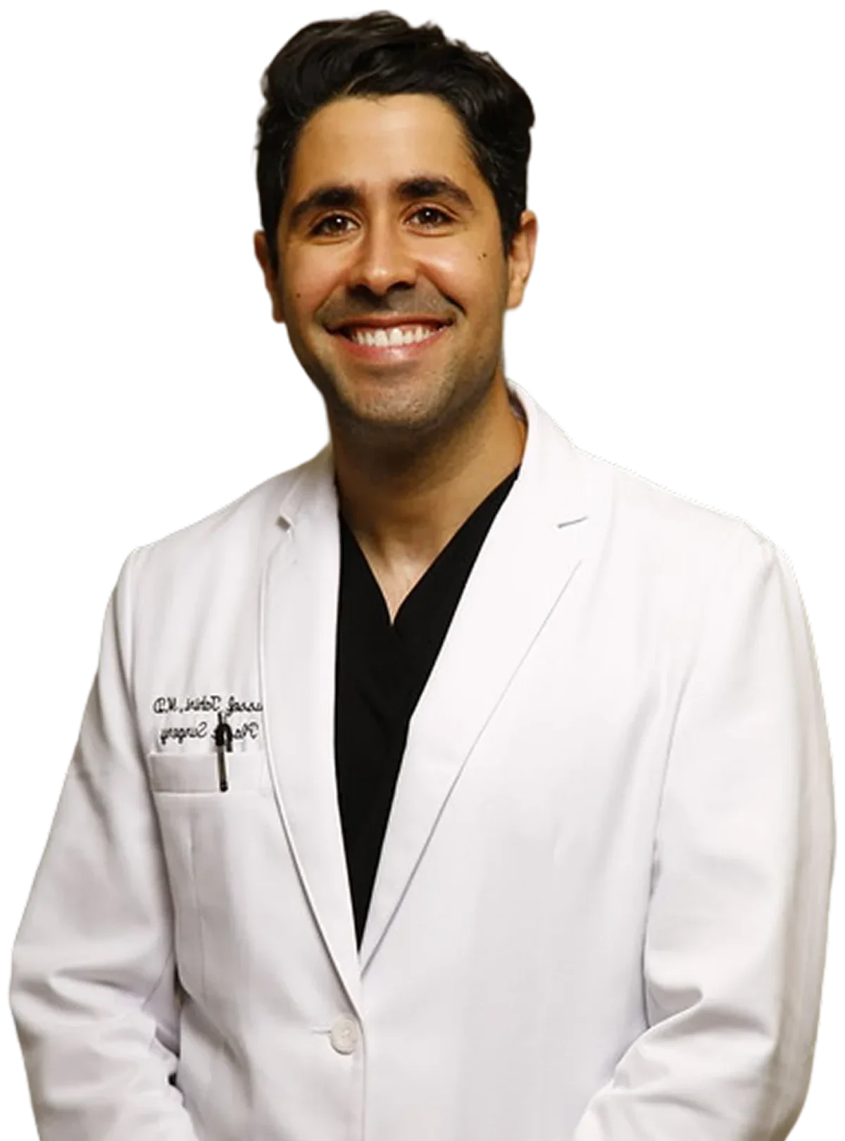
“We treat both congenital and acquired cases of marginal mandibular nerve palsy — evaluating the underlying cause to build a plan tailored to each patient’s age, anatomy, and goals.”
Dr. Tahiri, International Microtia Institute
Concerned about facial nerve involvement before or after a procedure? Dr. Tahiri’s team can walk you through your specific risk and options.
Schedule Your ConsultationPrevention and Risk Reduction:
What Is the Danger Zone for the Marginal Mandibular Nerve?
The marginal mandibular nerve sits near the lower border of your jaw, and its position makes it vulnerable during procedures in the neck, parotid, and submandibular regions.
In most people, the nerve runs within 2cm behind the corner of the mouth. Because of its superficial position here, surgeons often refer to this area as a “danger zone”—a region where even minor surgical errors or aggressive retraction can increase the risk of nerve injury.
Understanding this anatomy helps both patients and surgeons take the right precautions, reducing the likelihood of complications and protecting the delicate muscles that shape your lower lip movement and expressions.
Quick Reference: Surgical Safety
Danger Zone
- Lower jaw border
- ~2 cm behind mouth corner
- High-risk surgical region
Protection
- Landmark identification
- Layered dissection
- Nerve monitoring
- Fascial preservation
-
01 Nerve Protection
Anatomical Landmarks and Surgical Safety: How Do Surgeons Protect This Nerve During Operations?
When surgery takes place near your lower jaw or neck, your surgeon must work with precision because the marginal mandibular nerve sits close to the skin and can shift depending on your anatomy.
Protecting it requires both expertise and a deliberate step-by-step strategy.
Surgeons typically safeguard the nerve by using techniques such as:
- Accurate landmark identification: Your surgeon traces structures, such as the facial artery and the anterior facial vein, to understand where the nerve should be.
- Careful, layered dissection: Staying in the correct subplatysmal or fascial plane reduces the chance of cutting or stretching the nerve as tissues are separated.
- Sandwich technique: This creates a “protective envelope” of fascia and platysma around the nerve by dissecting above and below it, helping your surgeon avoid direct contact with the nerve.
- Node of Stahr approach: In this method, the surgeon identifies a specific lymph node as a landmark. They cut toward it until the marginal mandibular nerve becomes visible and can be safely tied off if needed.
- Hayes-Martin approach: The surgeon locates the facial vein and ties off two points, and makes a cut between them. The ends are then lifted upward to keep the nerve protected during the operation.
- Parotid-masseteric fascia approach: By following the fascia covering the parotid gland and masseter muscle, the surgeon uses this natural layer as a safe barrier, keeping them away from the nerve’s pathway.
- Pterygo-masseteric sling approach: This method involves cutting the masseter muscle to keep it out of the way, helping surgeons avoid the marginal mandibular nerve’s trajectory.
- Pouch technique: This technique involves making a small cut in the neck skin, opening it like a pouch, giving the surgeon open access to the underlying tissues while remaining mindful of the nerve’s position.
- Intraoperative nerve monitoring: Real-time metrics help your surgeon identify the nerve and confirm its function throughout the operation, especially during high-risk procedures.
With Dr. Tahiri, you have a surgeon who relies on meticulous technique and deep knowledge of facial anatomy.
His safety-focused approach, shaped by advanced craniofacial training and extensive experience, helps reduce the risk of nerve injury and leads to more predictable, confident surgical outcomes.
-
02 Safe Surgery
How Is Marginal Mandibular Nerve Palsy Prevented During Surgery?
Preventing marginal mandibular nerve palsy starts with a surgeon who understands exactly where the nerve travels and how its position can shift from patient to patient.
During procedures near the lower jaw, surgeons use several proven techniques to keep the nerve safe:
- Working in the subplatysmal plane to avoid the nerve’s most superficial segments. This layer allows surgeons to safely elevate tissue while protecting the nerve beneath the platysma.
- Identifying key anatomical landmarks, especially the facial artery, facial vein, the parotid-masseteric fascia, and the perifacial lymph nodes, which help map the expected path of the nerve and guide safe dissection.
- Recognizing the communication between the marginal mandibular and cervical branches of the facial nerve, which helps preserve function, even when individual anatomy varies.
- Using nerve monitoring or stimulation during higher-risk procedures to confirm the nerve’s location and avoid accidental stretching, compression, or transection.
- Applying nerve-preserving techniques such as the Hayes-Martin approach, where surgeons carefully manage the facial vein and surrounding fascia to maintain a safe distance from the nerve throughout the operation.
When you’re in the care of Dr. Youssef Tahiri, you benefit from a surgeon who blends advanced reconstructive training with meticulous technique and published anatomical expertise.
His deep understanding of facial nerve pathways—supported by medical research and textbook contributions—allows him to plan and perform surgery that minimizes nerve injury and protects natural lower lip function.
How Do You Treat Marginal Mandibular Nerve Palsy?
Treating marginal mandibular nerve palsy depends on the cause of the injury, how long the symptoms have been present, and how much function has been lost.
If the nerve is still intact, nonsurgical options may be enough to improve symmetry and movement.
However, if the nerve has been cut—or if meaningful improvement hasn’t returned within a reasonable timeframe—surgical reconstruction may offer the best results.
Treatment options range from simple balancing injections to advanced microsurgical procedures.
At International Microtia Institute, your treatment plan is tailored to your unique case. This personalized approach helps ensure that your recovery restores both natural movement and facial balance.
Will Marginal Mandibular Nerve Palsy Go Away on Its Own?
Whether your marginal mandibular nerve regains activity depends on the type of injury. If the nerve is just compressed or mildly stretched, you may see gradual improvement within a few weeks to several months.
If it’s a more serious injury, however, recovery may take longer, with some patients experiencing weakness of the lower lip and asymmetric smile for a year or longer.
Unfortunately, if a nerve is severed and becomes nonfunctional after an injury, it usually doesn’t recover on its own and often requires surgical repair.
Marginal mandibular nerve palsy does not behave like Bell’s palsy. Bell’s palsy often improves spontaneously because it affects the entire facial nerve and is usually linked to viral inflammation.
MMNP is more localized and less likely to recover without targeted treatment.
If you’re unsure which type of nerve injury you’re dealing with, Dr. Tahiri can evaluate your specific pattern of lower lip weakness, review your medical or surgical history, and help you understand whether natural recovery is likely or whether treatment may be needed for the best outcome.
RECOVERY & TREATMENT OPTIONS
What Recovery Can You Expect After a Marginal Mandibular Nerve Injury?
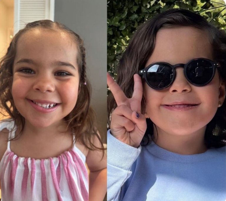
One of our happy patients before/after
A note from Dr. Tahiri & the care team
Every marginal mandibular nerve injury is different, and one of the first questions patients ask is, “Will this get better?” The answer depends on how the nerve was injured, how much of the nerve remains intact, and how soon treatment begins. Many patients with stretched or compressed nerves recover significant function over time—often without surgery. When the nerve has been completely severed, however, timely surgical repair offers the best opportunity for meaningful recovery. During your evaluation, Dr. Tahiri carefully assesses your nerve function, facial symmetry, and overall anatomy to explain what recovery is realistically possible and which treatment options are most appropriate for your situation.
What Is the Prognosis for Marginal Mandibular Nerve Injury?
Your prognosis depends on several factors, including how the injury occurred, how quickly treatment begins, and whether the nerve still has its structural continuity.
Compressed or stretched nerves typically have an excellent prognosis, often recovering full or near-full function within a few weeks or months. However, complete nerve transections (cutting or severing) require surgical repair for meaningful recovery.
Please note that the time since the injury, your age, and your individual anatomy all play a role in your overall outcome.
Modern anatomical classification systems also help predict which patients respond best to nerve repair, grafting, or muscle transfer.
With over 25,000 patients treated from more than 65 countries, Dr. Tahiri can assess your case with precision and outline a realistic prognosis based on your nerve function, facial symmetry, and clinical findings.
Nonsurgical Treatment Options
Nonsurgical options can help improve symmetry while the nerve heals or when surgery isn’t necessary. The two most popular are:
Chemodenervation with botulinum toxin injections on the unaffected side can balance lower lip movement. They soften the pull of the depressors and the platysma, temporarily improving symmetry.
Physical therapy and facial exercises may help maintain facial muscle function and reduce involuntary movements, especially during the nerve’s recovery phase.
These options can be effective when the nerve is expected to recover or when you prefer a nonsurgical approach. Dr. Tahiri will work with you to determine whether these options align with your goals and desired outcomes.
Surgical Treatment Options for Marginal Mandibular Nerve Injury
If the nerve is cut, severely damaged, or not recovering, surgery may restore natural lower lip movement. Fortunately, several options are available, including:
Quick Reference: Surgical Options
Repair
- Direct nerve repair
- Nerve grafting
Reanimation
- Muscle transfer
- Nerve transfer
Goal
- Restore movement
- Improve facial symmetry
-
01 Nerve Repair
Nerve Repair and Grafting
When the nerve ends are close enough, direct repair reconnects the marginal mandibular branch under a microscope. If there is a significant gap, a nerve graft bridges the distance.
These repairs work best when performed soon after the injury. Recovery can take several months as the nerve regenerates and reinnervates the depressor anguli oris and depressor labii inferioris.
An anatomical classification system helps determine whether direct repair or grafting is most likely to succeed.
In selected cases, direct muscle neurotization of the lower lip depressor muscles can be used to restore function.
-
02 Muscle Transfer
Muscle Transfer Procedures
Muscle transfer is an effective option when the nerve or muscle has lost long-term function.
Common approaches include:
- Anterior belly of digastric transfer: The digastric muscle is rerouted to recreate the downward pull of the lower lip.
- Free extensor digitorum brevis transfer: This procedure uses the extensor digitorum brevis (EDB) to offer reanimation of the lower lip muscles.
- Mylohyoid muscle transfer with tensor fascia lata graft: The mylohyoid muscle—innervated by a branch of the mandibular nerve—is sutured directly to the lower lip depressor muscles, often supported by a tensor fascia lata graft.
These procedures compensate for lost innervation and help restore natural facial expressions.
-
03 Nerve Transfer
Nerve Transfer Techniques
Nerve transfers reroute a functioning donor nerve to power the lower lip muscles. Options include:
- Mini-hypoglossal nerve transfer, which uses a small branch of the hypoglossal nerve as a motor donor to restore movement in lower lip muscles, including those with prolonged nonfunction.
- Cervical-to-marginal mandibular nerve transfer, where the cervical branch of the facial nerve is used as the donor nerve to reinnervate a nonfunctional marginal mandibular nerve.
Nerve transfers are especially useful for chronic injuries, longstanding paralysis, or cases where original nerve repair is not possible.
Advanced Surgical Techniques and Anatomical Considerations
Understanding the marginal mandibular nerve requires precision, especially when planning treatment for different levels of injury.
Surgeons classify these injuries into three zones because the location affects both outcomes and the techniques used. The levels are typically described as:
- Level 1 (Proximal): Injury is near or at the marginal mandibular nerve branch
- Level 2 (Parotid area): Injury is around the parotid area or in front of and below the ear
- Level 3 (Distal): Injury is far from the marginal mandibular nerve or is in the terminal branches
Each zone has its own challenges, which is why classification systems and quality-of-life assessments are essential during evaluation.
These levels guide decisions, help predict recovery, and ensure you receive an approach that delivers the most natural and functional result possible.
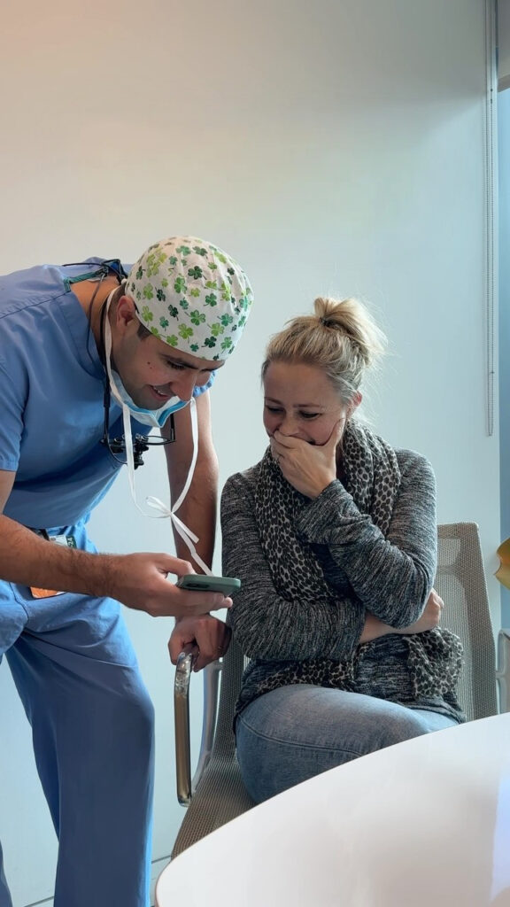
Anatomical Classification System for Treatment Planning
Surgeons rely on several grading tools to assess how your lower lip moves, the degree of weakness, and how the injury affects daily function.
These systems help compare pre- and postoperative results, keeping your treatment plan objective and tailored.
- Terzis’ lower lip grading scale evaluates symmetry and lower lip mobility.
- Photogrammetry uses standardized photographs to measure lip excursion and contour changes.
- Sunnybrook facial grading system provides a composite score based on symmetry at rest, voluntary movement, and synkinesis (involuntary muscle movement).
This standardized way of measuring facial function is described in a study published in Clinical Otolaryngology (Clin Otolaryngol. DOI: 10.1111). - Facial disability index measures how facial weakness affects everyday tasks and daily functioning.
- Facial clinimetric evaluation scale evaluates the functional and quality-of-life impact of facial nerve dysfunction.
Together, these classification systems guide the choice between nerve repair, nerve transfer, or muscle transfer so your treatment aligns with your functional needs and aesthetic goals.
Specialized Surgical Approaches
Some situations call for advanced reconstruction when the nerve cannot be repaired directly or when longstanding palsy affects muscle function.
Examples of these specialized surgical procedures include:
- Free gracilis muscle transplantation with microneurovascular anastomoses: The gracilis muscle is transplanted and connected to facial blood vessels and nerves to restore dynamic lower lip movement.
- Pectoralis minor vascularized muscle grafts: This procedure is designed for patients with facial paralysis who want to restore their ability to express emotions, such as laughter.
- Free microvascular and microneural transfer procedures: These techniques combine donor nerves and muscles to recreate coordinated movement, making it an excellent option for severe or complex cases of facial palsy.
Dr. Tahiri offers targeted procedures for mild to moderate asymmetry and more complex reconstruction for severe or longstanding cases, including:
- Selective myomectomy: A minimally invasive approach that weakens the DLI on the stronger, normally functioning side, which helps balance lower-lip movement and improve smile symmetry.
- Selective neurotomy: A procedure in which specific branches of the marginal mandibular nerve on the stronger side are partially divided to create a controlled, predictable improvement in symmetry.
Dr. Tahiri recommends these techniques selectively and thoughtfully, ensuring you receive the least invasive approach that can reliably restore balanced lower lip movement and long-term facial harmony.
Treatment Options for Different Patient Groups
Treatment for marginal mandibular nerve palsy isn’t one-size-fits-all. Every patient presents with a unique combination of age, anatomy, injury cause, and functional goals.
Dr. Tahiri, a renowned plastic surgeon who has treated children and adults from more than 65 countries, tailors each plan around these factors.
His expertise in both pediatric and adult craniofacial reconstruction allows him to adjust timing, technique, and long-term planning so each patient receives the safest and most effective solution for their situation.
Quick Reference: Find Your Path
Children
- Congenital conditions
- Growth-aware treatment
Cancer & Trauma
- Reconstruction planning
- Coordinated care
Surgical Injuries
- Personalized treatment
- Repair or rehabilitation
-
01 Pediatric Care
Treatment for Children and Congenital Cases
Children with congenital or early-onset marginal mandibular nerve dysfunction require a different treatment pathway because their facial structures are still growing and developing.
Here are some considerations:
- Timing of intervention: Early evaluation is important, but surgery is often timed carefully to avoid disrupting natural facial growth.
- Some congenital cases improve as the child grows, while others require staged reconstruction once facial proportions become more stable.
- Growth-related concerns: Treatment must account for ongoing mandibular and soft-tissue development.
- Procedures such as nerve or muscle transfers are designed with the child’s future growth in mind to ensure harmonious results over time.
- Family counseling and long-term planning: Parents should be guided through expected outcomes, developmental milestones, and functional goals such as feeding, speech, and emotional expression.
With advanced training in pediatric craniofacial surgery, rest assured that Dr. Tahiri can help your child with marginal mandibular nerve palsy.
His approach considers symmetry, psychosocial development, and the need for age-appropriate intervention strategies.
-
02 Cancer & Trauma
Treatment After Cancer Surgery or Trauma
When the marginal mandibular nerve is intentionally sacrificed during cancer surgery or damaged during an accident, treatment must be coordinated carefully based on timing, tissue quality, and overall health.
- Reconstructive timing: Some patients benefit from immediate nerve repair or grafting at the time of tumor removal or trauma repair.
Others require delayed reconstruction once oncologic treatment is complete and the surgical site has healed. - Coordination with oncology and reconstruction teams: For patients recovering from ablative cancer procedures, Dr. Tahiri works alongside oncology specialists to ensure reconstruction does not interfere with cancer surveillance or radiation therapy planning.
Please understand, however, that injuries in the digastric region often involve deeper structures and may require more advanced treatments, such as nerve repair, nerve transfers, or muscle transfer techniques.
For this reason, Dr. Tahiri selects the most appropriate approach based on your unique situation—including the remaining nerve length, muscle viability, and expected recovery potential.
- Reconstructive timing: Some patients benefit from immediate nerve repair or grafting at the time of tumor removal or trauma repair.
-
03 Post-Surgical Care
Treatment Considerations for Different Surgical Causes
The optimal treatment approach depends greatly on how the nerve was injured and the type of surgery involved. In simple terms, different procedures carry different risks and leave varying reconstructive options.
- After parotidectomy (parotid gland removal): Parotid gland surgery—especially when performed to remove carcinoma—can lead to facial paralysis if the facial nerve is sacrificed during the procedure.
- After submandibular gland excision: Injuries in this region typically affect the marginal mandibular nerve, with a PubMed report showing an approximately 15.6% incidence rate.
- After neck dissection: Neck dissections often damage the marginal mandibular nerve. That said, severe injuries and facial asymmetry following a neck dissection are relatively rare.
- After carotid endarterectomy (carotid artery plaque removal): The marginal mandibular branch is the second most frequently affected during this procedure, with an incidence rate as high as 27%.
Depending on how the injury occurred, recovery may occur spontaneously, or surgical intervention may be required if lower lip movement does not return.
Across all surgical causes, Dr. Tahiri evaluates the level of injury, the timing, and the specific functional deficits to choose the most effective treatment plan—whether that involves observation, focused rehabilitation, or advanced reconstructive surgery designed to restore natural facial expression and symmetry.
What to Expect
During Recovery and Follow-Up
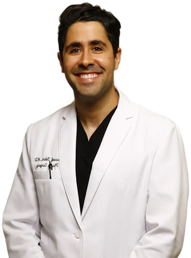
Dr. Tahiri, International Microtia Institute
Recovery from marginal mandibular nerve palsy depends on the cause of the injury and the treatment performed.
Whether your care plan involves nonsurgical therapy or reconstructive surgery, Dr. Tahiri provides a structured follow-up plan so you understand what’s normal, what to watch for, and when improvement is likely to occur.
This follow-up period includes nerve-healing timelines, muscle recovery, and individualized care instructions based on your age, anatomy, and overall health.
Note that neurophysiological factors, such as axonal regrowth speed, synaptic reinnervation, and muscle readiness, influence the pace and completeness of recovery.
Your follow-up schedule typically includes early wound checks, nerve function assessments at key intervals, and long-term monitoring to assess symmetry, lip movement, and overall facial balance.
How Long Does Recovery Take?
Recovery is highly dependent on the type of nerve injury and the procedure performed. Below is a realistic, medically accurate timeline to help set expectations.
If the nerve was only compressed or mildly stretched (neuropraxia):
- Improvement may begin within weeks.
- Most patients recover fully within a few months.
- Children often recover faster due to stronger neuroplasticity.
If the nerve was significantly stretched (axonotmesis):
- Nerve fibers regenerate at roughly 1mm per day (one inch per month).
- Recovery typically takes five to six months.
- Some residual synkinesis may persist, depending on the degree of injury.
If the nerve was cut (neurotmesis):
- Surgical repair must occur within two to six weeks after the initial injury.
- Visible functional improvement usually appears around three months.
- Full recovery may take up to two years, depending on the type of repair and the muscle’s responsiveness.
When it comes to recovery after facial paralysis or nerve injury, outcomes fall along a spectrum, from full restoration to lasting changes, depending on the injury’s severity and how quickly it’s treated.
In a nutshell, mild damage typically heals completely on its own. Moderate injuries usually improve with time, but subtle signs may linger.
Severe or untreated nerve transections, on the other hand, are different. If the nerve is severed and left unrepaired for too long, muscles begin to permanently waste away.
Without surgical intervention, the paralysis on that side of the face might remain permanent—resulting in no movement, a flattened expression, or functional issues like a drooping mouth.
What Are the Risks and Complications?
All treatments—whether nonsurgical or surgical—carry some degree of risk. Here are the potential risks and complications associated with treating marginal mandibular nerve palsy (MMNP):
Nonsurgical Treatment Risks
- Botulinum toxin injections: temporary weakness in nearby muscles and need for repeat injections every three to four months.
- Physical therapy: mild soreness or limited improvement if the underlying nerve is severely damaged.
Surgical Treatment Risks
Nerve repair or grafting
- Incomplete recovery
- Scar formation
- Synkinesis (unwanted movement during smiling or other expressions)
Muscle transfer procedures
- Donor-site morbidity
- Bulkiness or contour irregularities
- Delayed reinnervation or limited return of movement
Nerve transfers
- Weakness in the muscle used as the donor nerve source
- Risk of incomplete reinnervation
- Need for neurological adaptation as patients learn to activate the new muscle pathway
General Surgical Risks
- Infection
- Hematoma (blood collection)
- Unfavorable scarring
- Wound healing delays
- Rare complications related to anesthesia
Fortunately, Dr. Tahiri takes a safety-first approach, informed by experience treating more than 25,000 patients. His meticulous technique and compassionate, education-focused care help minimize complications and support predictable, stable outcomes.
Under Dr. Tahiri’s care, most patients experience a smooth recovery, steady improvement, and long-lasting results.
Choosing the Right Surgeon
Selecting the right surgeon is one of the most important decisions you will make when dealing with marginal mandibular nerve palsy.
Because this nerve is small, delicate, and highly variable in its anatomy, successful treatment requires a surgeon who not only understands facial nerve pathways but has extensive experience. Here’s what truly matters when choosing a surgeon for this condition:
-
Deep understanding of facial anatomy: Your surgeon should have precise knowledge of the marginal mandibular nerve’s location, its variations, and its relationship to surrounding tissues.
-
Expertise in facial nerve procedures: Treating this nerve requires specialized surgical skills and an eye for choosing the optimal approach for each patient.
-
Advanced fellowship training: Surgeons with dedicated craniofacial or facial nerve reconstruction training are better equipped to handle complex cases.
-
Experience supported by thousands of cases: Outcomes improve significantly with high surgical volume. Dr. Youssef Tahiri has treated over 25,000 patients from more than 65 countries, giving him one of the broadest and most diverse clinical experiences.
-
Published medical authority and textbook co-authorship: Dr. Tahiri has contributed to the scientific literature and co-authored respected surgical textbooks. This academic foundation reinforces his role as a thought leader in craniofacial and facial nerve surgery, translating into safer planning, improved precision, and consistently better results.
-
Commitment to communication and personalized care: Dr. Tahiri is thorough, honest, and attentive—qualities that matter when recovering from a nerve injury and navigating your treatment options.
Choosing a surgeon with proven expertise minimizes your risk, improves your chances of functional recovery, and ensures you receive a clear, thoughtful, and compassionate treatment roadmap.
Dr. Tahiri’s blend of high-level training, international reputation, and surgical excellence makes him uniquely qualified to treat both simple and highly complex marginal mandibular nerve injuries.
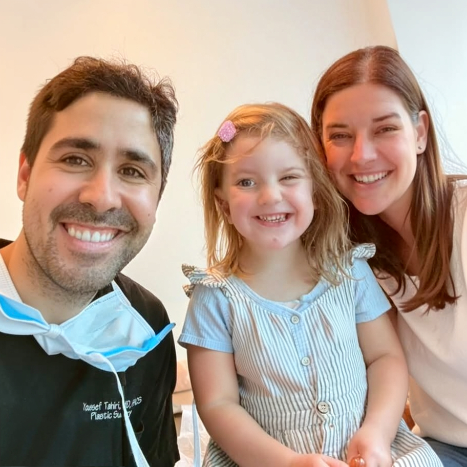
When to Seek Treatment from Dr. Tahiri
If you’re noticing lower lip asymmetry, difficulty forming certain sounds, or weakness that appeared after surgery or trauma, it may be time to consult a specialist who understands the full spectrum of MMNP.
You should also seek evaluation if the problem has been present for months without improvement or if you’re unsure whether the nerve is bruised, stretched, or permanently damaged.
Dr. Youssef Tahiri is uniquely positioned to help you at any stage.
He treats both acute injuries—where early intervention may restore nerve function—and chronic cases, where advanced reconstruction can improve symmetry and movement.
His expertise spans both pediatric and adult patients, supported by his craniofacial surgery background and high-volume experience treating more than 25,000 patients from over 65 countries.
Whether you live locally or are traveling from abroad, his Beverly Hills practice offers seamless support for national and international patients.
Dr. Tahiri provides undivided attention, clear communication, and a best-care promise that prioritizes your long-term function and appearance.
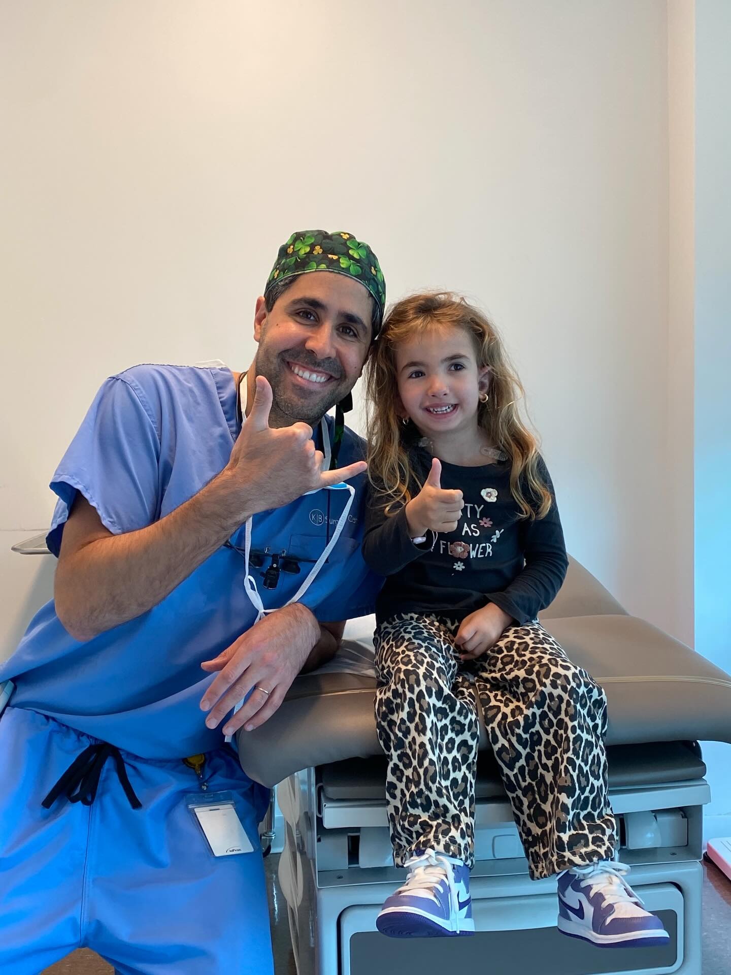
What Makes Dr. Tahiri’s Approach Different?
Dr. Youssef Tahiri’s treatment philosophy is grounded in precision, compassion, and world-class surgical training.
His background includes advanced specialization in plastic, reconstructive, and craniofacial surgery, allowing him to treat even the most complex cases with confidence and accuracy.
What sets his approach apart:
- Expertise in complex craniofacial cases: He routinely treats nerve injuries, whether marginal mandibular nerve palsy or congenital conditions like hemifacial microsomia.
- Mastery of microsurgical and reconstructive techniques: Dr. Tahiri offers muscle- and nerve-focused balancing procedures for facial paralysis.
- Published authority and textbook co-author: His academic contributions ensure you’re receiving care from a surgeon recognized by peers for his advanced knowledge and expertise.
- Global reputation: Patients from 65+ countries travel to him for his surgical outcomes, meticulous planning, and commitment to patient-centered care.
- Specialization in SUPOR™ and craniofacial reconstruction: His skill with delicate anatomical structures gives him a precision advantage in lower facial surgery.
By combining technical excellence with transparent communication, Dr. Tahiri ensures you receive thoughtful guidance tailored to your specific condition.
Testimonials
What our Patients Say About Us
-
What an incredible experience with Dr Tahiri! He has great bedside manners, his clinic had excellent staff and our son had a great result with his ear reconstruction. I would highly recommend him to any parents looking for a plastic surgeon to perform their child’s microtia reconstruction.

-
Dr. Tahiri is an exceptional plastic surgeon. I have referred many friends to Dr. Tahiri for their own problems as well as their childrens’! He is renowned for his microtia ear reconstructions but his results in all plastic surgery procedures are flawless! He is such a talented surgeon with amazing bedside manners, communication style, an amazing team that makes you feel comfortable at all times. Thank you Dr. Tahiri!
Julian

-
Dr Tahiri is great. Great surgeon. Great guy. He made me feel at ease, encouraged questions and dialogue, but most of all performed incredibly.
I went to him for reconstruction on my nose after having a basal cell removed. A very tricky and intricate procedure that could’ve altered my appearance.
It was and is perfection.
I highly recommend him if you need a delicate hand and superior craftsmanship.

-
He is the best Dr I have ever seen when dealing with children. He made our daughter so comfortable. We are so happy we were referred to him.

-
An amazing surgeon, after about six microtia surgeries that did not result the way I wanted them to, I decided to go for the medpor, it was a scary decision to make because I was afraid that it would turn out just like the rest of my previous surgeries but what happened was that I got an ear that I am happy to show off to everyone, and I find it great that he was able to work off of the damage from my previous surgeries and still make it look so nice, I can finally pull my hair into ponytails without feeling insecure and its all thanks to Dr. Tahiri

-
Dr. Tahiri is an exceptionally gifted and talented surgeon. He performed my reconstructive jaw surgery to correct my class 3 malocclusion—something I had been wanting to correct for the past decade. From the first appointment, I knew that he was the surgeon I wanted to complete this major procedure because he made me feel comfortable and confident in his skills as a surgeon, especially when discussing the complexities of my case. He is incredibly personable, sincere, bright, and invested in his patient’s well-being; his enthusiasm and smile are contagious! Furthermore, his staff at Cedars-Sinai are some of the most professional and caring healthcare providers I have ever met. This surgery changed my life for the better and I will forever be grateful for this excellent doctor and team.

-
Amazing Surgeon did excellent with my daughter in her microtia repair. Very kind and invested in making a difference in these childrens lives. Was very supportive to me as a mother as I we went through this process would not chose any other surgeon.

-
Dr. Tahiri is a professional in every way. Every visit is an unforgettable experience.
She is a kind and warm person with a wonderful approach to children.
We have been under his care for several years and we recommend everyone to meet such a man and doctor on their path.
His team are wonderful people always helpful in any situation. We hope that also this time the visit will bring the desired result. For everything from the bottom of our hearts thank you.
-
Dr Tahiri is great! He’s very kind and easy to talk to. My daughter had a dermoid cyst above her eyebrow that Dr Tahiri removed at Cedars. Now, a year later, the scar is not even visible. We are very happy we found him and would highly recommend him.

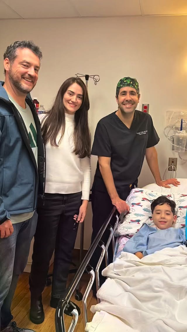
Schedule a Consultation with Dr. Tahiri
If you’re ready to understand your options and get clarity about your condition, scheduling a consultation is the next step.
You’ll meet directly with Dr. Tahiri, who takes the time to listen, evaluate your nerve function, and explain every treatment path in detail.
During your visit, you’ll receive:
- A personalized treatment plan built around your goals
- Expert craniofacial and facial nerve evaluation
- Clear explanations of surgical and nonsurgical options
- Access to patient testimonials and video success stories that show real outcomes
Whether your case is new or longstanding, Dr. Tahiri will guide you with unmatched precision and a genuine commitment to helping you achieve the best possible result.
Schedule your consultation today by calling (310) 255-4476 or emailing [email protected].
Your Questions Answered
Frequently Asked Questions About Marginal Mandibular Nerve Palsy
How much will marginal mandibular nerve palsy bother someone?
Marginal mandibular nerve palsy can affect people differently, but it commonly impacts both daily function and emotional well-being.
- Functional issues: Difficulty pulling the lower lip down can affect speech, eating, and saliva control.
- Cosmetic concerns: Lower lip asymmetry becomes noticeable when making facial expressions.
- Emotional impact: Even mild asymmetry can make someone feel self-conscious or less expressive.
The good news is that treatment—whether nonsurgical or surgical—can often restore movement, improve symmetry, and help you or your loved one feel more comfortable and confident.
What precautions are necessary with marginal mandibular nerve palsy?
Taking the right precautions can help protect the affected area, prevent symptoms from worsening, and support long-term recovery.
Here are some key daily care tips to keep in mind:
- Avoid excessive stretching or pressure:
Your body will naturally try to compensate for the weakness over time. Until then, be mindful of avoiding accidental biting or stretching of the lower lip, as it may be more vulnerable on the weaker side. - Seek immediate medical attention if you experience:
- A rapid onset of new facial weakness
- Severe swelling, bruising, or signs of infection
- Difficulty eating or speaking that worsens quickly
- Patient counseling considerations:
Everyone heals differently. A consultation with a facial nerve specialist can help you understand what changes to expect, how facial movement may evolve, and what treatment timelines are realistic for your situation.
With the right guidance and support, many people find that thoughtful care and early intervention make a meaningful difference in their recovery.
Does marginal mandibular neuropathy improve over time?
Many cases do improve spontaneously. That said, several factors influence natural improvement, including:
- Type of injury: Mild nerve injuries heal on their own within weeks to months. More severe injuries, however, may not recover fully without surgical intervention.
- Age-related anatomy: Younger patients typically have better regenerative potential. If you’re older, healing may happen more slowly, and recovery may be less complete without treatment.
- Nerve branch communication: The marginal mandibular nerve has variable connections with nearby facial nerve branches, including the anterior and buccal branches.
Stronger cross-communication between these branches can improve spontaneous recovery and help the face compensate more effectively.
What is the difference between temporary and permanent nerve damage?
Understanding whether nerve damage is temporary or permanent is essential for setting expectations and choosing the right treatment plan.
Temporary nerve damage:
- Lower lip weakness gradually improves over weeks to months.
- Sensation and movement may return fully.
- Can be treated with nonsurgical options like BOTOX® to support symmetry during recovery.
Permanent nerve damage:
- Often caused by surgery-related injury
- The lower lip may remain asymmetrical without intervention
- Requires surgical repair or reanimation procedures to restore symmetry and movement
How clinicians distinguish between the two:
- EMG (electromyography): Helps assess whether the nerve is still sending signals.
- Tracking improvement over time: Continued progress suggests a temporary injury.
- Intraoperative nerve assessment: Used in certain cases to evaluate nerve integrity directly.
Knowing which type of injury you’re dealing with helps you and your surgeon make informed decisions about treatment and long-term outcomes.
How are surgical outcomes measured?
Evaluating outcomes is essential—not just to confirm that function and appearance have improved, but also to give you clarity and reassurance during the healing process.
When results feel uncertain, it’s completely natural to start researching on your own, sometimes diving into Google Scholar in search of answers. A clear, structured way of measuring progress can help replace that anxiety with confidence.
Here are some of the most common tools and methods surgeons use to assess outcomes:
- House–Brackmann scale: A widely used system for grading facial nerve function. It evaluates symmetry, movement, and muscle tone on a scale from I (normal) to VI (complete paralysis).
- Photogrammetry: Standardized before-and-after photography allows precise measurement of lip position, symmetry, and dynamic movement. This allows surgeons to track even subtle improvements over time.
- Video-based movement analysis: Pre- and post-op video recordings help evaluate lower lip movement, coordination, and expression patterns.
- Clinical physical examination: Surgeons assess muscle tone, voluntary movement, compensatory mechanisms, and synkinesis.
- Patient-reported outcomes: Self-assessments of confidence, comfort, and functional improvement offer valuable insight.
Research published in Plastic and Reconstructive Surgery—Global Open (Plast Reconstr Surg Glob Open. DOI: 10.1097) highlights the importance of these patient experiences.
Together, these tools help surgeons quantify progress and ensure that treatment restores not only aesthetic balance but also meaningful facial function.
Contact Dr. Tahiri
Get in Touch
Choosing a surgeon for you or your child is one of the biggest decisions you’ll ever make. You want someone with the right training, the right experience, and the right approach.
Dr. Tahiri checks all those boxes. But more than that, he understands what this journey means for your family. He knows that behind every medical case is a child with dreams and parents who want the very best for them.
You don’t have to figure this out alone. We’re here to help, and we’re ready when you are.
Call the Office (310) 255-4476“Amazing experience with Dr Tahiri and his team. Everyone made us feel so comfortable and safe and eased all my fears of having my 5 year old go through surgery… “
Contact Dr. Tahiri today by filling out the form above or calling the office at (310) 255-4476. For families outside the Los Angeles area: We’re happy to start with a virtual consultation to discuss you or your child’s condition and determine if an in-person visit makes sense.
Dr. Tahiri accepts most PPO insurance plans. Our team will guide you through every step of the process, from initial consultation to complete recovery.